
- Home Page
- Company Profile
-
Our Products
- Histopathology Products
- Microtomes
- ConXport ADVANCE ROTARY MICROTOME
- Automatic Cryostat Microtome
- ConXport Automatic Cryostat Microtome
- ConXport . Hand & Table Microtome
- ConXport Semi Automatic Rotary Microtome
- Freezing Microtome
- ConXport Fully Automatic Rotary Microtome
- Fully Automatic Microtome
- ConXport Advance Rotary Manual Microtome
- ConXport Freezing Microtome
- ConXport Rotary Microtome (New Model)
- ConXport ROTARY MICROTOME SPENCER
- Erma Microtome
- ConXport Advance Rotary Microtome
- ConXport Manual Rotary Microtome
- ConXport Fully Automated Microtome Touch Screen
- ConXport Semi-Automatic Microtome Touch model
- ConXport Rocking Microtome
- SEMI AUTOMATIC MICROTOME
- ConXport Rotary Microtome (New Model)
- ConXport Automatic Knife Sharpener For Microtome
- ConXport Senior Precision Rotary Microtome
- ConXport Rotary Microtome
- ConXport . Sliding / Sledge Microtome
- ConXport Sliding Microtome
- ConXport Semi Automatic Rotary Microtome
- Advance Rotary Microtome
- Advance Rotary Manual Microtome
- Freezing Microtome
- Rotary Microtomespencer
- Rocking Microtome
- Rotary Microtome
- Rotary Microtome (New Model)
- Senior Precision Rotary Microtome
- Sliding Microtome
- ConXport Manual Rotary Microtome
- Pharmacy Equipment
- ConXport . Dissolution Single Test Apparatus
- ConXport . Du-Bios Reymond Key
- ConXport . Disintegration Single
- ConXport Ampoule Filling & Sealing Machine
- ConXport Tablet Making Machine Hand Operated
- ConXport . Antibiotic Zone Reader
- ConXport Double Cone Blender
- ConXport Pohl Commutator
- ConXport . Pulverizer Machine
- Lab Stirrer
- Jar Test Apparatus
- Disintegration Test Apparatus Tablet
- Motorized Tablet Making Machine
- Hand Operated Tablet Making Machine
- Pharmacy Capsule Filling Machine
- Hand Operated Bottle Sealing Machine
- Tablet Disintegration Double Test Apparatus
- ConXport Disintegration Test Apparatus Tablet
- ConXport Tablet Making Machine Motorized
- ConXport . Dissolution Six Test Apparatus
- ConXport Pharmacy Capsule Filling Machine
- ConXport Tablet Disintegration Double Test Apparatus
- ConXport . Bottle Sealing Machine Hand Operated
- ConXport Ampoule Clarity Test Apparatus
- ConXport . Electric Tablet Friability Test Apparatus Single Drum
- ConXport Ampoule Washing Machine
- ConXport Bulk Density Apparatus
- ConXport . Perimeter Priestley Smith
- ConXport Glass Bead Sterilizer
- ConXport . Jar testing Apparatus Spindle Stirrer
- ConXport . Student Organ Bath
- ConXport Student Organ Bath Double Unit
- ConXport . Hammer Mill
- ConXport JAR TEST DIGITAL LCD DISPLAY
- ConXport . Vortex Shaker
- ConXport . All Purpose Equipment
- ConXport . Mechanical Stirrer
- ConXport . Dales Organ Bath
- ConXport Digital Kymograph
- ConXport Simple Electrode
- ConXport Collapsible Tube Filling Machine
- ConXport . Collapsible Tube Sealing Machine
- ConXport CAPSOULE FILLING MACHINE
- ConXport Analgesiometer (Eddy's Hot Plate)
- ConXport . Collapsible Tube Filling
- ConXport Student Spirometer
- ConXport Tablet Hardness Tester Pfizer
- ConXport TABLET HARDNESS TESTER (MONSANTO TYPE)
- ConXport . Skin Fold Caliper
- ConXport Suppository Mould
- ConXport Bottle Washing Machine
- ConXport Glass Bead Sterilizer
- ConXport Piston Recorder
- ConXport Swimming Test Apparatus
- ConXport Elevated Plus Maze
- ConXport Tablet Coating Pan
- ConXport Tissue Culture Station
- ConXport Sterility Test Apparatus
- ConXport Vibrating Interrupter
- ConXport . Polishing Pan
- ConXport . Tincture Press
- ConXport . Student-Kymograph-Recording-Drum
- ConXport . BOTTLE FILLING MACHINE HAND OPERATED
- ConXport . Rota Rod 4 Comp
- ConXport Electro Convulsometer
- ConXport Ball Mill
- ConXport Super Speed Kymograph
- ConXport . Dissolution Single Digital R.P.M. & Temp.
- ConXport Bottle Filling Motorized Machine
- ConXport Bicycle Ergometer
- ConXport Tablet Dissolution Test Apparatus
- ConXport . Rota Rod 2 Comp.
- ConXport . Glassware Dryers
- ConXport Tissue c
- ConXport . Actophotometer
- ConXport . Mammalian Heart Perfusion Assembly
- ConXport Rota Rod 4 Comp.
- ConXport . Friability Double
- ConXport . Analgesiometer Tail Flick
- ConXport Vibrating Reed
- Rotary Vacuum Evaporator
- Covid-19 Products
- ConXport Fogging Machine
- Sanitizer Stand
- ConXport Electric Steam Vaporizer 3 in 1
- Conxports Stainless Steel Sanitizer Stand
- ConXport Safety Goggles
- ConXport 3 Layer Mask With Nose Pin
- ConXport N95 Mask
- ConXport Shoe Cover
- ConXport Disposable non-woven Bouffant Cap
- ConXport Hand Sanitizer 5ltr
- ConXport Nebuliser
- ConXport Sodium Hypochlorite
- ConXport 100% Polyester Swab Stick
- ConXport VTM Kit
- ConXport Laboratory Thermometer
- Ambulance And Recovery Equipment
- ConXport Autoloader Collapsible Stretcher (Ambulance Stretcher)
- ConXport Ambulance Stretcher Cum Wheelchair
- ConXport Wheelchair Stair Stretcher
- ConXport Folding Stretcher
- ConXport Folding Stretcher 2 Fold Aluminium
- ConXport Folding Stretcher 2 Fold Mild Steel
- ConXport Folding Stretcher 4 Fold
- ConXport Basket Stretcher
- ConXport Head Immobilizer
- ConXport Spine Board
- ConXport 2 Folded Spine Board
- ConXport Scoop Stretcher Telescopic
- ConXport Plastic Scoop Stretcher
- ConXport Adult Traction Splint Set
- ConXport Air Splint Set of 6
- ConXport Rolling Splint Adjustable
- ConXport Emergency Blanket Gold & Silver
- ConXport Emergency Blanket Gold & Silver
- ConXport Carry Sheet Blue
- ConXport Cervical Collar Adjustable
- ConXport Extraction Device
- ConXport Oxygen Cylinder Aluminium 1 Ltr.
- ConXport Oxygen Cylinder Aluminium 2ltr
- ConXport Oxygen Cylinder Aluminium 3 ltr
- ConXport Oxygen Cylinder Aluminium 5ltr
- ConXport Oxygen Cylinder Aluminium
- ConXport Hand Held Suction Device
- ConXport Vacuum Stretcher
- ConXport Patient Transfer Stretcher
- ConXport Emergency Scissor
- Anesthesia Equipment
- ConXport Oropharyngeal Guedel Airways Set of 8
- ConXport CPR Mask Pocket Type Silicone
- ConXport Anaesthesia Machine
- Conxport Anaesthesia Machine Portable
- Conxport Laryngoscope Faibre Optic Miller 4 Blades Ss Plastic Box
- Conxport Anaesthesia Machine
- ConXport Black Rubber Face Mask
- ConXport Oxygen Mask with Reservoir Bag
- ConXport Silicone Face Mask
- ConXport Oropharyngeal Guedel Airways Pink
- ConXport Oropharyngeal Guedel Airways Blue
- ConXport Oropharyngeal Guedel Airways Black
- ConXport Oropharyngeal Guedel Airways White
- ConXport Oropharyngeal Guedel Airways Green
- ConXport Oropharyngeal Guedel Airways Yellow
- ConXport Oropharyngeal Guedel Airways Red
- ConXport Oropharyngeal Guedel Airways Sky Blue
- ConXport Nasopharyngeal Airways
- Conxport Anesthesia Workstation
- Conxport Laryngeal Mask Airway Pvc
- ConXport Laryngeal Mask Silicone Single
- ConXport Laryngeal Mask Silicone Reusable
- ConXport Anaesthesia Face Mask
- ConXport Antistatic Face Mask
- ConXport CPR Mask Child Silicone
- ConXport CPR Mask Adult Silicone
- ConXport Oxygen Mask
- ConXport Rendell Bracker Face Mask
- ConXport Rendell Bracker Face Mask Silicone
- ConXport Venturi Mask
- ConXport CPAP Full Face Mask
- ConXport NIV Mask
- ConXport Silicone Resuscitator Infant / Ambu Bag
- ConXport Silicone Resuscitator Infant With Airways / Ambu Bag
- ConXport Silicone Resuscitator Child / Ambu Bag
- ConXport Silicone Resuscitator Child / Ambu Bag With Airways
- ConXport Silicone Resuscitator Adult or Ambu Bag
- ConXport Silicone Resuscitator Adult or Ambu Bag With Airways
- ConXport Black Rubber Resuscitator Infant / Ambu Bag
- ConXport Black Rubber Resuscitator Child or Ambu Bag
- ConXport Black Rubber Resuscitator Adult or Ambu Bag
- Conxport Laryngoscope Fibre Optic Miller 3 Blades Ss Rexine Pouch
- ConXport Laryngoscope Fibre Optic Miller 3 Blades SS Plastic Box
- ConXport Laryngoscope Fibre Optic Miller 4 Blades SS Rexine Pouch
- ConXport Laryngoscope Set Miller Stainless Steel 3 Blades
- ConXport Laryngoscope Set Miller Stainless Steel 3 Blades Plastic Box
- ConXport Laryngoscope Set Miller Stainless Steel 4 Blades Rexine Pouch
- Conxport Laryngoscope Set Miller Stainless Steel 4 Blades Plastic Box
- ConXport Ventilator Portable
- ConXport Ventilator Vertical
- ConXport Oxygen Concentrator Single Flow 5 Ltr
- ConXport Oxygen Concentrator Single Flow 10 Ltr
- ConXport Oxygen Concentrator Dual Flow
- ConXport Oxygen Cylinder Mild Steel
- ConXport Nitrogen Cylinder Or Nitrous Oxide Cylinder
- ConXport Oxygen Cylinder Aluminium 5 Ltr
- ConXport Oxygen Cylinder Aluminium 10 Ltr
- ConXport Cylinder Trolley U Handle
- ConXport Cylinder Trolley H Handle
- ConXport Medical Spanner Stainless Steel
- ConXport Medical Cylinder Key
- ConXport Oxygen Cylinder Regulator
- ConXport Nitrous Oxide Regulator
- ConXport Mox Regulator
- ConXport Oxygen Regulator with Flow Meter and Humidifier Bottle
- ConXport Fine Adjustment Valve Rota Meter and Humidifier Bottle
- ConXport Humidifier Bottle
- ConXport T Oxygen Recovery Kit
- ConXport Oxygen Nasal Cannula
- Autopsy Equipment
- ConXport Autopsy Table Stainless Steel
- ConXport Autopsy Table Electro Hydraulic
- ConXport Autopsy Forensic Workstation
- ConXport Dead Body Bags Center Zipper
- ConXport Dead Body Bags Army
- ConXport Dead Body Transfer Trolley
- ConXport Dead Body Wash Trolley
- ConXport Mortuary Trolley Height Adjustable
- ConXport Dead Body Concealment Trolley or Morgue Trolley
- ConXport Dead Body Weighing Scale
- ConXport Organ Weighing Scale
- ConXport Embalming Machine Mobile
- ConXport Embalming Machine Table Top
- ConXport Embalming Workstation
- ConXport Oscillating Autopsy Saw
- ConXport Autopsy Saw With Vacuum Dust Collector
- ConXport Anthropometric Rod
- ConXport Headrest Plastic Six Position
- ConXport Mail Gloves SS Chain
- ConXport Osteometric Board
- ConXport Post Mortem Set
- ConXport Silicone Oil For Post Mortem Instrument
- ConXport Mortuary Cabinet 1 Compartment
- ConXport Mortuary Cabinet 2 Compartment
- ConXport Mortuary Cabinet 3 Compartment
- ConXport Mortuary Cabinet 4 Compartment
- ConXport Mortuary Cabinet 6 Compartment
- ConXport Dead Body Freezer
- ConXport Hydraulic Dead Body Lifter
- ConXport Electro Hydraulic Dead Body Lifter
- Infant Care Equipment
- Baby Bassinet With Utility Box
- Infant Radiant Warmer
- ConXport Open Care System
- ConXport Baby Bassinet With Hydraulic
- ConXport Baby Bassinet Standard
- ConXport Baby Bassinet with Plastic Crib
- ConXport Baby Bassinet Luxury
- ConXport Baby Infant Incubator
- ConXport Baby Infant Incubator With Phototherapy
- Baby Transport Incubator Standard
- Overhead Phototherapy Unit LED
- Overhead Phototherapy Unit CFL
- Double Surface Phototherapy Unit CFL
- Double Surface Phototherapy Unit LED
- Undersurface Phototherapy Unit CFL
- Undersurface Phototherapy Unit LED With Wheel
- ConXport Penguin Sucker
- ConXport Jaundice Meter or Bilirubinometer
- Digital Infant Resuscitator
- ConXport Infant Resuscitator with Oxygen Blender
- ConXport Infant Trolley With Resuscitator
- ConXport Infant Trolley Without Resuscitator
- ConXport Fetoscope Plastic
- ConXport Fetoscope Wooden
- ConXport Fetoscope Aluminium
- ConXport Fetoscope Stethoscope
- ConXport Infant Vein Viewer
- ConXport Pencil Vein Viewer
- ConXport Infrared Vein Finder Multi Purpose
- ConXport C Vein Finder
- ConXport Oxygen Hood With Nozzle
- ConXport Fetal Heart Doppler
- ConXport Bubble Cpap System
- ConXport Baby Bath Tub Round Shape
- ConXport Baby Bath Tub Rectangular Shape
- ConXport Baby Bottle Plastic
- Blood Bank Equipments
- ConXport Safety Sampling System Triple
- ConXport TRANSFER BAG
- ConXport Quadruple Leukocyte Reduction Filter
- ConXport Blood Bag Tube Sealer With Battery Backup
- ConXport BLOOD BAG ICE BOX WITH DISPLAY
- ConXport BLOOD BANK REFRIGERATED CENTRIFUGE
- ConXport Blood Transfusions Chair
- ConXport Blood Bag Tube Sealer Without Battery Backup
- ConXport Plasma Extractor Manual
- ConXport Double Pan Balance / Scale
- ConXport Plasma Extractor Electric
- ConXport Blood Donor Camp Chair
- ConXport Blood Bag Tube Hand Sealer
- ConXport Blood Donors Chair
- ConXport Electric Blood Donors Chair / Dialysis Chair
- ConXport Blood Donor Chair Economy
- ConXport Blood Bag Quadruple Cod / Sag
- ConXport Platelet Agitator
- ConXport Cryon Bath
- ConXport BLOOD BANK REFRIGERATOR
- ConXport Cooling Incubator
- ConXport Blood Bag Quadruple Cpda-1
- ConXport Infusion Fluid Warmer
- ConXport Blood Collection Monitor With Battery Backup
- ConXport Blood Donor Couch 3 Functions Electric
- ConXport Single Pan Balance / Scale
- ConXport Blood Donor Couch 2 Functions Electric
- ConXport Blood Bag Triple Cod / Sag
- ConXport Platelet Incubator
- ConXport Blood Roller Mixer
- ConXport Plasma Thawing Bath
- ConXport Blood Bag Single Cpda-1
- ConXport Infusion Fluid Warmer Double Line
- Conxport Blood Collection Monitor Without Battery Backup
- ConXport Blood Bag Double Cpda-1
- ConXport Blood Bag Triple Cpda-1
- ConXport Blood Bank Refrigerator Glass Door
- Diagnostic Products
- ConXport Head Band Mirror Set
- ConXport Head Light Focused German Type
- ConXport Aneroid Bp Cuff Adult
- Conxport Vaginal Speculum Medline Breisky
- Conxport Queen Square Hammer
- ConXport Bp Machine Digital Wrist Model Citizen Ch 650
- Diagnostic Audiometer
- Conxport Stethoscope Diaphragm
- ConXport Clark Head Light
- Conxport Nebulizer Omron C 25s
- Conxport Otoscope Welch Allyn 22870 Blk
- Conxport Magnifier With Tennis Racket
- Conxport Glucometer Accu Chek Active Strips
- Conxport Watchmaker Glass
- Conxport Stethoscope Frame
- Conxport Stethoscope Ear Piece
- Conxport Magnifier With Bakelite Handle
- Conxport Magnifier With Metal Handle
- Conxport Nebulizer Mask
- Conxport Thread Counter Metal
- Conxport Stethoscope Tubes
- ConXport Ent Set Welch Allyn Set Of 2
- Conxport Glucometer Dr. Morepen Strips
- ConXport Magnifier Tripod
- Conxport Mouth Examination Mirrors
- Conxport Pocket Magnifier Metal
- Conxport Otoscope Welch Allyn 20000
- Conxport Vaginal Speculum Sims
- Conxport Vaginal Speculum Doyen
- Conxport Vaginal Speculum Graves
- Conxport Vaginal Speculum Plastic
- ConXport Head Light Focused German Type With Transformer
- ConXport Bp Machine Led Mercury Free
- Conxport Vaginal Speculum Kristeller
- Conxport Tongue Depressor Wooden Sterile
- ConXport Bp Machine Lcd Mercury Free
- Conxport Thermometer Mercury Deluxe
- Conxport Tongue Depressor Wooden Non Sterile
- Conxport Otoscope Welch Allyn 22840
- Conxport Percussion Hammer Pin Brush
- Conxport Stethoscope Teaching
- ConXport Otoscope Welch Allyn 23920
- Conxport Stethoscope Dual Head Adult
- Conxport Stethoscope Single Head Adult
- ConXport Ent Set Welch Allyn 777
- ConXport Bp Machine Mobile Mercury Ms Base
- Conxport Stethoscope Cardio
- ConXport Ear Syringe Metal
- ConXport Bp Machine Mercury Wall Mounted
- ConXport Bp Machine Bulb Green
- ConXport Bp Machine Bulb Black
- Conxport Ear Syringe Glass
- Conxport Stethoscope Fetoscope
- ConXport Bp Machine Digital Automatic Citizen Ch 432
- Conxport Otoscope Welch Allyn 22860
- Conxport Bp Machine Digital Automatic Omron 7124
- ConXport Ear Syringe Plastic
- ConXport Bp Machine Digital Wrist Model Citizen Ch 650
- Conxport Tongue Depressor Stainless Steel L Shape
- ConXport Glucometer Dr Morepen With 25 Strips
- Conxport Stethoscope Dual Head Paediatric
- ConXport Bp Machine Metal Valve
- Conxport Thermometer Mercury Economy
- Concxport Laryngeal Mirrors
- ConXport Bp Armlet Bag
- Conxport Nebulizer Omron C 28
- Conxport Vaginal Speculum Auvard
- Conxport Tuning Fork Stainless Steel
- Conxport Magnifier With Light
- ConXport BP MACHINE MERCURY DESK
- Conxport Neurological Hammer
- Conxport Thread Counter Plastic
- Conxport Tongue Depressor Stainless Steel S Shape
- ConXport Ear Speculum
- Conxport Nebulizer Omron C 803
- ConXport Clark Head Light With Transformer
- ConXport Head Light Led
- Conxport Ophthalmoscope
- Conxport Thermometer Forehead
- ConXport Ent Diagnostic Set Basic
- ConXport Ear Syringe Rubber
- Conxport Nebulizer Nulife Handyneb
- Conxport Otoscope
- Conxport Neurological Hammer Pin Brush
- ConXport Glucometer Accu Chek Active
- ConXport Bp Machine Mobile Mercury Pu Base
- Conxport Otoscope Welch Allyn 23820
- Digital Audiometer Nano
- Diagnostic Audiometer
- Conxport Berliner Percussion Hammer
- ConXport Vaginal Speculum Collin
- Conxport Stethoscope Cardiology
- Conxport Vaginal Speculum Cuscos
- Hospital Furniture
- ConXport Automatic Tissue Processor
- Grossing Station
- ConXport Paraffin Dispensers
- ConXport Slide Warming Table
- ConXport . Cryostat Microtome Semi Automatic
- ConXport Burn Cage
- ConXport Paraffin Wax Trimmer
- ConXport Linear Slide Stainer
- ConXport Tissue Flotation Bath
- ConXport . Microprocessor Tissue Processor
- ConXport Cryostat Microtome Manual
- ConXport Plain Bed Standard Laminated Panel with Wheels
- ConXport Cooling Plate
- ConXport Slide Staining Machine
- ConXport . Vacuum Tissue Processor
- ConXport Grossing Workstation Single person
- Dental Chair
- ICU Bed
- ConXport Icu Bed Electric With Central Locking
- ConXport ICU Bed Electric Collapsible Railings
- ConXport ICU Bed Pediatric Electric With Railings
- ConXport ICU Bed Electric ABS Panels & ABS Railings
- Conxport ICU Bed Electric SS Panels & Collapsible Railings
- ConXport ICU Bed Electric with ABS Top
- ConXport ICU Bed Semi Electric
- ConXport Full Fowler Bed Electric ABS Top
- ConXport Full Fowler Bed Electric
- ConXport Semi Fowler Bed Electric ABS Top
- ConXport Plain Bed Standard with Wheels
- ConXport Plain Bed Wire Mesh Top with Wheels
- ConXport Plain Bed Wire Mesh Top Laminated Panel with Wheels
- ConXport Orthopaedic Bed ABS Panels
- ConXport Electric Delivery Bed
- ConXport Hydraulic Delivery Bed
- ConXport Manual Delivery Bed 2 Section
- ConXport Manual Delivery Bed 3 Section
- ConXport Delivery Couch 3 Section
- ConXport Telescopic Delivery Obstetric Bed
- ConXport Obstetric Labour Table Mechanical
- ConXport Telescopic Labour Table Fixed Height
- ConXport Telescopic Labour Table Adjustable Height
- ConXport Gynae Table Plain
- ConXport Gynae Table 2 Section
- ConXport Gynae Couch With Cabinet
- ConXport Instrument Trolley 2 Shelves Standard
- ConXport Instrument Treatment Trolley ABS 2 Shelves
- ConXport Dressing Trolley Standard
- ConXport Dressing Trolley with Drawer
- ConXport Mayo Instrument Trolley Standard
- ConXport Mayo Instrument Trolley Deluxe
- ConXport Mayo Instrument Trolley with Gear Handle
- ConXport Medicine Trolley 2 Drawer 1 Shelve
- ConXport Medicine Trolley with Bowl & Tray
- ConXport Medicine Trolley with Five Drawer
- ConXport Medicine Trolley with Six Drawer
- ConXport Stretcher Trolley Metal Top
- ConXport Stretcher Trolley Stainless Steel
- ConXport Stretcher Trolley with Canvas Top
- ConXport Stretcher Trolley Folding
- ConXport Stretcher Trolley with Mattress
- ConXport Patient Transfer Trolley System
- ConXport Patient Trolley 2 Section with Crank
- ConXport Emergency and Recovery Trolley Hydraulic
- ConXport Bedside Locker Standard Plain Top
- ConXport Open Bedside Locker
- ConXport Bedside Locker ABS
- ConXport Bedside Locker with Overbed Table
- ConXport IV Stand Double Hook PU Base
- ConXport IV Stand Double Hook Metal Base 3 Wheels
- ConXport IV Stand Double Hook Metal Base 4 Wheels
- ConXport IV Stand Double Hook Metal Base 5 Wheels
- ConXport IV Stand Four Foldable Hook PU Base
- ConXport IV Stand Four Foldable Hook Metal Base 3 Wheels
- ConXport IV Stand Four Foldable Hook Metal Base 4 Wheels
- ConXport IV Stand Four Foldable Hook Metal Base 5 Wheels
- ConXport Bowl Stand Single Metal Base
- ConXport Bowl Stand Single with Frame 3 Legs
- ConXport Bowl Stand Single with Frame 4 Legs
- ConXport Bowl Stand Double Metal Base
- ConXport Bowl Stand Double with Frame 3 Legs
- ConXport Bowl Stand Double with Frame 4 Legs
- ConXport Drum Stand Single
- ConXport Drum Stand Double
- ConXport Baby Cradle
- ConXport Baby Cradle Foldable
- ConXport Infant Pediatric Cot
- ConXport Revolving Stool Cushion Top
- ConXport Revolving Stool SS Top
- ConXport Revolving Stool SS Top 3 Legs
- ConXport Visitor Stool Fixed
- ConXport Revolving Stool Cushion with Wheels
- ConXport Revolving Stool SS with Wheels
- ConXport Revolving Stool with Footrest
- ConXport Surgeon Stool Revolving
- ConXport Surgeon Stool Revolving Deluxe
- ConXport Surgeon Chair with Arm & Foot Rest
- ConXport Ambulatory Chair
- ConXport Bedside Screen 2 Panels
- ConXport Bedside Screen 3 Panels
- ConXport Bedside Screen 4 Panels
- ConXport Electric Blood Donor Chair / Dialysis Chair
- ConXport Blood Transfusion Chair
- ConXport Blood Donor Chair
- ConXport Waiting Chair Plastic 4 Seater
- ConXport Waiting Chair Plastic 2 Seater
- ConXport Waiting Chair Plastic 3 Seater
- ConXport Waiting Chair Plastic 5 Seater
- ConXport Waiting Chair Metal 2 Seater
- ConXport Waiting Chair Metal 3 Seater
- ConXport Overbed Bedside Table Fixed Sunmica Top
- ConXport Overbed Bedside Table Adjustable Manual
- ConXport Overbed Bedside Table Adjustable Gear Handle
- ConXport Overbed Table ABS
- ConXport Overbed Bedside Table Adjustable 2 Section
- ConXport Plaster Table
- ConXport Orthopaedic Plaster Table
- ConXport Swaddling Table Baby Table
- ConXport Kick Bowl 3 Legs
- ConXport Kick Bowl 4 Legs
- ConXport Kick Bucket Standard
- ConXport Kick Bucket Deluxe
- ConXport First Aid Cabinets
- ConXport Filing Cabinets 2 Drawer
- ConXport Filing Cabinets 3 Drawer
- ConXport Filing Cabinets 4 Drawer
- ConXport Instrument Cabinet One Door
- ConXport Instrument Cabinet Three Door
- ConXport Instrument Cabinet Four Door
- ConXport Storage & Instrument Cabinet Metal
- ConXport Storage & Instrument Cabinet Glass Door
- ConXport Foot Step Rubber Top Single
- ConXport Foot Step Rubber Top Double
- ConXport Foot Step Metal Top Single
- ConXport Foot Step Metal Top Double
- ConXport Foot Step Laminated Top Single
- ConXport Foot Step Laminated Top Double
- ConXport Foot Step ABS Top Single
- ConXport Foot Step ABS Top Double
- ConXport Anaesthesia Trolley ABS
- ConXport Case History Trolley Single
- ConXport Case History Trolley Double
- ConXport Monitor Trolley
- ConXport Patient Record Trolley
- ConXport Utility Trolley One Drawer
- ConXport Utility Trolley Two Drawer
- ConXport Crash Cart Trolley Mild Steel
- ConXport Crash Cart Trolley Stainless Steel
- ConXport Soiled Linen Trolley Canvas Bag Round
- ConXport Soiled Linen Trolley Canvas Bag Square
- ConXport Soiled Linen Trolley Plastic Bucket
- ConXport Linen Change Trolley
- ConXport Soiled Linen Trolley Square
- ConXport Oxygen Cylinder Trolley Small U Handle
- ConXport Oxygen Cylinder Trolley Small H Handle
- ConXport Waste Bin Plastic with Closed Lid
- ConXport Waste Bin Plastic with Flap Lid
- ConXport Waste Bin with Wheels
- ConXport Waste Bin Color Coded 32L
- ConXport Waste Bin Metal Plain
- ConXport Waste Bin Metal Plain with Pedal
- ConXport Medical Waste Bin Metal with Wheels
- ConXport Waste Bin Metal Perforated
- ConXport Waste Bin Metal Perforated with Pedal
- ConXport Waste Bin Metal with Flap Lid
- ConXport Feminine Sanitary Bin PP
- ConXport Mattress Plain
- ConXport Mattress Two Section
- ConXport Mattress Four Section
- ConXport Mattress Five Section
- ConXport Air Mattress Anti Decubitus
- ConXport Air Mattress Tube Type
- ConXport Backrest Canvas
- ConXport Electric Backrest
- ConXport Case Sheet Holder
- ConXport Case Sheet Holder Blue
- ConXport Balkan Beam Frame
- ConXport Side Railing Aluminium
- ConXport Side Railing Stainless Steel
- ConXport Side Railing ABS
- ConXport ABS Head and Foot Board Economy
- ConXport ABS Head and Foot Board Deluxe
- ConXport ABS Head and Foot Board Compound
- ConXport ABS Head and Foot Board with Bumper
- ConXport Baby Crib
- ConXport Dining Board Adjustable
- ConXport Lifting Pole
- ConXport Mosquito Net Pole
- ConXport 4 Bed Pan 4 Urinal Rack
- ConXport 6 Bed Pan 6 Urinal Rack
- ConXport Urinal Rack
- ConXport Bowl & Basin Rack
- ConXport Open Rack
- ConXport Shoes Rack
- ConXport Swab Rack
- ConXport Bed Elevator
- ConXport Air Cushion
- ConXport Rubber Bed Pan
- ConXport Breast Pump Plastic
- ConXport Breast Pump Plastic With Container
- ConXport Breast Pump Plastic With Bottle
- ConXport Breast Pump Glass
- ConXport Chloroform Bellow
- ConXport Corrugated Rubber Drainage Sheet
- ConXport Rubber Douche Bag 1 Ltr
- ConXport Rubber Douche Bag 2 Ltr
- ConXport Rubber Douche Bag 4 Ltr
- ConXport Douche Fitting Set
- ConXport Douche Tube
- ConXport Rubber Water Bottle Both Side Ribbed
- ConXport Rubber Water Bottle Both Side Ribbed With Handle
- Plain Bed
- HD Virtual Dissection Table 88
- 3d Virtual Dissection Table Mini
- Virtual Dissection Table Anatovizz
- Hospital Hollowares
- ConXport Urinal Female Plastic Non Autoclavable
- ConXport Kidney Tray Plastic Economy
- ConXport Mayo Tray S/S 202 Grade
- ConXport Dressing Drum Jointed S/S 202 Grade
- ConXport Thermometer Jar S/S 202 Grade
- ConXport Baby Tray Plastic
- ConXport Instrument Tray With Cover S/S 202 Grade Economy
- ConXport Instrument Tray Without Cover S/S 202 Grade Deluxe
- ConXport Kidney Tray With Cover S/S 304 Grade
- ConXport Kidney Tray Without Cover S/S 304 Grade
- ConXpert Douche Can SS 202 Grade
- Aluminium Surgical Instrument Box
- ConXport Mayo Tray S/S 304 Grade
- SS Surgical Instrument Box
- SS Bucket
- ConXport Bed Pan Male Plastic Non Autoclavable
- ConXport BED PAN FEMALE PLASTIC AUTOCLAVABLE
- ConXport Urinal Female S/S 304 Grade
- ConXport URINAL FEMALE PLASTIC AUTOCLAVABLE
- ConXport Bed Pan Female S/S 304 Grade Seamless
- ConXport Bed Pan Female Plastic Non Autoclavable
- ConXport Bed Pan Male S/S 202 Grade Seamless
- ConXport Douche Can Tube
- Green Color Plastic Bowl
- ConXport BED PAN MALE S/S 304 GRADE SEAMLESS
- ConXport Douche Can Fitting Set
- ConXport Kidney Tray Without Cover S/S 202 Grade
- SS Round Gallipot Without Cover
- Plastic Surgical Basin
- ConXport Bed Pan Male Plastic Autoclavable
- ConXport Bed Pan Male S/S 202 Grade Jointed
- Stainless Steel Surgical Bowl
- SS High Quality Instrument Box
- ConXport Bed Pan Female S/S 202 Grade Jointed
- ConXport Urinal Female S/S 202 Grade
- ConXport Thermometer Jar Plastic
- ConXport URINAL MALE S/S 202 GRADE
- ConXport Urinal Male Plastic Autoclavable
- ConXport Bed Pan Female Round S/S 202 Grade Seamless
- ConXport Measuring Jar S/S 202 Grade
- ConXport Bed Pan Female S/S 304 Grade Jointed
- ConXport Bed Pan Male S/S 304 Grade Jointed
- SS Round Bucket With Handle
- White Plastic Bucket
- Round Cylindrical Shape Gallipot Without Cover
- SS Round Basin
- SS Gallipot With Cover
- ConXport Forceps Jar Plastic
- ConXport Thermometer Jar S/S 304 Grade
- ConXport Measuring Jar Plastic
- ConXport Douche Can Plastic
- SS Surgical Sterilizer Box
- SS Hospital Bowl Cover
- SS Round Gallipot With Cover
- SS Surgical Bowl Without Cover
- ConXport Urinal Male S/S 304 Grade
- ConXport Bed Pan Female Without Cover Seamless
- ConXport Urinal Male Plastic Non Autoclavable
- ConXpert Douche Can SS 304 Grade
- SS Wash Basin
- ConXport Cidex Tray S/S 202 Grade
- ConXport Kidney Tray Plastic Deluxe
- ConXport Kidney Tray Without Cover S/S 304 Grade
- ConXport Instrument Tray Plastic
- ConXport Instrument Tray With Cover S/S 304 Grade
- ConXport Kidney Tray Plastic Deluxe With Handle
- ConXport Dressing Drum Jointless S/S 202 Grade
- ConXport Catheter Tray With Cover S/S 202 Grade
- ConXport Kidney Tray With Cover S/S 202 Grade
- ConXport Instrument Tray Aluminium
- ConXport Shallow Tray S/S 202 Grade
- ConXport Forceps Jar S/S 202 Grade
- ConXport Instrument Tray With Cover S/S 304 Grade
- ConXport Catheter Tray With Cover S/S 304 Grade
- ConXport Instrument Tray With Cover S/S 202 Grade Deluxe
- ConXport Baby Tray S/S 202 Grade
- ConXport Shallow Tray S/S 304 Grade
- ConXport Dressing Drum Jointless S/S 304 Grade
- ConXport Dressing Drum Jointed S/S 304 Grade
- ConXport Cidex Tray Plastic
- ConXport Cidex Tray S/S 304 Grade
- ConXport Baby Tray S/S 304 Grade
- ConXport Instrument Tray Without Cover S/S 202 Grade economy
- SS Surgical Bowl Cover
- ConXport Dressing Jar S/S 202 Grade
- ConXport Bed Pan Female S/S 202 Grade Seamless
- Weight And Measuring Scale
- Conxport Bathroom Scale 130 Kg Round
- Conxport Measuring Tapes 5 Mtr
- Conxport Baby Weighing Scale Hanging Tubular Aluminium
- ConXport Height Measuring Rod Wall Mounted
- ConXport Bathroom Scale 150 Kg Round Manufactured By Conxport
- ConXport Bathroom Scale 150 Kg Bmi
- ConXport Kitchen Scales Round Top
- Conxport Baby Weighing Scale Hanging Dial Type
- Conxport Analytical Balances With Internal Calibration
- Conxport Baby Weighing Hammock
- Conxport Baby Weighing Trousers
- Conxport Circular Weighing Scale
- Conxport Bathroom Scale 130 Kg Square
- Conxport Baby Scale Digital 20 Kg 10 Gms
- ConXport Kitchen Scales Square Top
- ConXport Diet Scale With Bowl
- ConXport Bathroom Scale 150 Kg Square
- Conxport Measuring Tapes 3 Mtr
- Conxport Measuring Tapes 30 Mtr
- ConXport Bathroom Scale Digital 150 Kg Square Glass
- ConXport Multipurpose Weighing Scale 10 Kg
- ConXport Multipurpose Weighing Scale 2 Kg
- Conxport Platform Balances
- Conxport Measuring Tapes 15 Mtr
- ConXport Height Measure With Digital Weighing Scale
- ConXport Weighing Scales Mechanical Column Type
- Conxport Table Top Digital Scale
- ConXport Baby Weighing Scale Hanging Tubular Nylon
- ConXport Infantometer Acrylic
- Conxport Hanging Tubular Scale Acrylic
- ConXportDigital 150 Kg Round Glass
- ConXport Infant Scale Plastic Tray
- ConXport Infant Height Measuring Scale Wooden
- ConXport Multi Colored Measuring Tape 1.5 Mtr
- ConXport Stature Meter
- Conxport Infant Scale Metal Base
- Conxport Measuring Tapes 10 Mtr
- Conxport Baby Weighing Scale Hanging Salter Plastic
- ConXport Height Measuring Rod For Babies
- ConXport Infant Scale Plastic Base
- Conxport Multi Colored Measuring Tape 2 Mtr
- ConXport Lab Scale Round Top
- ConXport Height Measure With Round Base
- ConXport Height Measure With Mechanical Weighing Scale
- ConXport Weighing Scales Mechanical Dial Type
- Conxport Baby Cum Child Scale
- ConXportWeighing Scales Digital
- Conxport Bathroom Scale 130 Kg Round With Handle
- Conxport Baby Weighing Scale Hanging Salter Metal
- Conxport Analytical Balances With External Calibration
- ICU Products
- ConXport Veterinary Multipara Monitor
- ConXport PULSE OXIMETER NEONATAL
- ConXport Bi-Clamp Cable Cord
- ConXport 5 Parameter Patient Monitor 10"
- ConXport Ecg Electrode Rectangular Shape
- ConXport Fetal Monitor 8.4" Display
- ConXport Needle Set
- ConXport Monopolar Active Handle Cable Cord
- ConXport Laparoscopy Bi-Clamp (Vessel Sealer)
- ConXport Patient Plate Cable Cord
- ConXport Bipolar Forceps Cable Cord
- ConXport Single Paddle Foot Switch
- ConXport Ecg Roll 12 Channel
- ConXport Bipolar Forceps
- ConXport Hand Switch
- ConXport BIPOLAR FORCEPS REGULAR BAYONET
- ConXport Ecg Gel
- ConXport Silicone Plates
- ConXport Ecg Roll 6 Channel
- ConXport Ecg Roll 3 Channel
- ConXport Surgical Monopolar Active Handle Valley Lab Type
- ConXport Set Of 3 Electrodes
- ConXport Patient Plate S/S
- ConXport Ecg Roll 1 Channel
- ConXport Double Paddle Foot Switch
- ConXport Hand Switch Pencil Disposable
- ConXport Fogger Mechanical
- ConXport Cautery Machine 300 Watt Digital
- ConXport Radio Frequency Cautery
- ConXport Cervical Cautery
- ConXport Halogen Light Source
- ConXport Dvt Pump
- ConXport Electro Skin Cautery
- ConXport Vessel Sealer With Surgical Cautery In Built (Digital)
- ConXport Spirometer Desktop
- ConXport Cautery Machine 300 Watt Analog
- ConXport Defibrillator Aed
- ConXport Ecg Machine 12 Channel With Usb
- ConXport Cautery Machine 400 Watt Analog
- ConXport Etco2 Monitor
- ConXport Led Light Source
- ConXport Auto Cpap Machine
- ConXport Fetal Monitor 7" Display
- ConXport Fetal Monitor 10.1" Display
- ConXport Defibrillator Monitor Aed
- ConXport Volumetric Infusion Pump
- ConXport Cautery Machine 400 Watt Digital 99 Programmable
- ConXport Ecg Electrode Oval Shape
- ConXport Spirometer Diagnostic
- ConXport Fogger Digital
- ConXport B-Pap Machine
- ConXport Co2 Insuffulator
- ConXport Syringe Pump
- ConXport Volumetric Infusion Pump Large Display
- ConXport CAUTERY MACHINE 400 WATT DIGITAL
- ConXport Defibrillator Monitor Only Ecg
- ConXport Defibrillator Monitor With Ecg & Printer
- ConXport Syringe Pump Colour Display
- ConXport Ecg Machine 12 Channel
- ConXport 5 Parameter Patient Monitor 15"
- ConXport Ecg Machine 1 Channel
- ConXport 5 Parameter Patient Monitor 12.1"
- ConXport Veterinary Pulse Oximeter
- ConXport PULSE OXIMETER HANDHELD
- ConXport Ecg Machine 3 Channel
- ConXport ECG MACHINE 6 CHANNEL WITH TOUCH SCREEN
- ConXport Ultrasound Scanner Black And White
- ConXport 5 Parameter Patient Monitor 8.4"
- ConXport Ecg Electrode Round Shape
- Hospital Disposables
- ConXport Tracheostomy Tube Plain
- ConXport 3 Way Stop Cock
- ConXport Infusion Set Deluxe
- ConXport Stitch Cutter
- ConXport Surgical Blades Stainless Steel
- ConXport BLOOD TRANSFUSION SET
- ConXport 3 Way Stop Cock With Extension Line
- ConXport Manual Vacuum Aspiration Kit
- ConXport Skin Stapler Remover
- ConXport Unolock Syringe Without Needle
- ConXport Delivery Kit
- ConXport Endotracheal Tube Holder
- ConXport Surgical Blades Carbon Steel
- ConXport Disposable Scalpel Handle
- ConXport Cytobrush / Endocervical Brush
- ConXport Pen Needle
- ConXport Disposable Scalpel Handle Thumb Type
- ConXport Spinal Needle Pencil Point
- ConXport High Pressure Monitoring Line
- ConXport Silicone Foley Balloon Catheter 2 Way Adult Bh
- ConXport Mucus Extractor
- ConXport Mr Syringe Kit
- ConXport Silicone Foley Balloon Catheter 3 Way Adult Bh
- ConXport Under Water Sealed Drainage System
- ConXport Intubating Stylette
- ConXport Infusion Set With Builtin Airvent
- ConXport Paul s Tracheal Culture Bottle
- ConXport Umbilical TAPE
- ConXport Spinal Needle Quincke Point
- ConXport Silicone Foley Balloon Catheter 2 Way Paed
- ConXport Urine Collection Bag Paediatric
- ConXport Umbilical Cord Clamp
- ConXport Dispovan Syringe Without Needle
- ConXport Infusion Set Economy
- ConXport Dispovan Tuberculin Syringe
- ConXport Infant Feeding Tube With Graduation
- ConXport Abdominal Drainage Kit
- ConXport Burette Set (Measured Volume Paediatric Set)
- ConXport Closed Wound Suction Unit
- ConXport Silicone Foley Balloon Catheter 2 Way Paed Bh
- ConXport Male Circumcision Kit
- ConXport Foley Balloon Cathterization Kit
- ConXport Chest Drainage Bag
- ConXport Urine Collection Bag Top Outlet
- ConXport Salem Sump Tube
- ConXport Infant Feeding Tube
- ConXport Silicone Foley Balloon Catheter 3 Way Adult
- ConXport Scalp Vein Set
- ConXport Microdrip Infusion Set
- ConXport Dispovan Needles
- ConXport Dispovan Insulin Syringe
- ConXport Pressure Monitoring Line
- ConXport Skin Stapler Regular
- ConXport Urine Collection Bag With Measured Volume Meter
- ConXport Foley Balloon Catheter 2 Way High Flow Adult
- ConXport Foley Balloon Catheter 3 Way Adult
- ConXport Intravenous Cannula
- ConXport Skin Stapler Wide
- ConXport Colostomy Bag
- ConXport Medical Disposable Renal Dilator Set
- ConXport Urine Collection Bag Top Outlet With Handle
- ConXport Infusion Set Standard
- ConXport Foley Balloon Catheter 3 Way High Flow Adult
- ConXport Foley Balloon Catheter 2 Way High Flow Paed
- ConXport Endotracheal Tube Cuffed South Oral Preformed
- ConXport Endotracheal Tube Subglottic Suction Line
- ConXport Endotracheal Tube Plain South Oral Preformed
- ConXport Endotracheal Tube Cuffed Pvc
- ConXport Endotracheal Tube Plain North Nasal Preformed
- ConXport Endotracheal Tube Cuffed North Nasal Preformed
- ConXport Foley Balloon Catheter 2 Way Paed
- ConXport Endotracheal Tube Plain Reinforced
- ConXport Foley Balloon Catheter 2 Way Adult
- ConXport Endotracheal Tube Plain Elastomer Coated
- ConXport Endotracheal Tube Cuffed Elastomer Coated
- ConXport Endotracheal Tubes
- ConXport Endotracheal Tube Cuffed Reinforced
- ConXport BOUGIES VENTED
- ConXport Silicone Foley Balloon Catheter 2 Way Adult
- ConXport Tracheostomy Tube Cuffed
- X-Ray Products
- ConXport X-Ray Processing Tank
- ConXport X-Ray Gonad Shield
- ConXport X-Ray Face Shield Mask
- ConXport X-Ray Dark Room Safe Light
- ConXport X-Ray Lead Goggles Metal Frame
- ConXport X Ray View Box Single With Sensor and Dimming Function
- ConXport X Ray Ceiling Suspension Shield With OT Light
- ConXport X Ray Cassette Screen Blue
- ConXport X Ray View Box Single Slim Digital With Sensor and Dimming Function
- ConXport X Ray View Box Single Slim Digital With Sensor
- ConXport x ray shield grids
- ConXport X-Ray Ovary Shield
- ConXport X-Ray View Box Triple Slim Digital With Sensor
- ConXport X-RAY LEAD LINED PROTECTION SCREEN HALF
- ConXport Ultrasound Gel Blue
- ConXport X-Ray Double Sided Apron Without Sleeves
- ConXport X-Ray Cassette Screen Green
- ConXport X-Ray Jacket
- ConXport X-Ray View Box Quadruple With Sensor
- ConXport X-Ray H.T. Cable
- ConXport Open Bi-Clamp
- ConXport X-Ray Arm Guard
- ConXport X-Ray Lead Number
- ConXport X-Ray Film Holding Clip
- ConXport X-Ray Lead Lined Protection Screen Full
- ConXport Pulse Oximeter Desktop With Nibp Lcd Display
- ConXport X-Ray Bucky
- ConXport X-Ray View Box Double With Sensor
- ConXport X-Ray Collimeter
- ConXport X-Ray Table Mounted Shield
- ConXport X-Ray View Box Quadruple Slim Digital With Sensor and Dimming Function
- ConXport X-Ray Cassette Holder Chest Stand Platform Model
- ConXport X-Ray Lead Goggles Metal Frame with Side Shields
- ConXport X-Ray Envelopes
- ConXport Lead Sheett
- ConXport X Ray Stationary Tube
- ConXport X-Ray Cassette Film Cart
- ConXport X-Ray Lead Glass
- ConXport X-Ray Lead Goggles Wrap-Around
- ConXport X-Ray Film Storage Box
- ConXport X-Ray Leg Guard
- ConXport X-Ray Protection Suspension Ceiling
- Conxport X-ray Lead Gloves
- ConXport X-Ray Lead Cap
- ConXport X-Ray Lead Lined Protection Screen Adjustable
- ConXport Ultrasound Gel Transparent
- ConXport x-ray film hanger
- ConXport X-Ray Fixer
- ConXport Ultrasound Scanner Color
- ConXport X-Ray Rotating Tube
- ConXport Automatic Film Processors Compact-2
- ConXport X-Ray Lead Goggles Full View
- ConXport X-Ray Developing Tank
- ConXport X-RAY VIEW BOX TRIPLE WITH SENSOR
- ConXport Automatic Film Processors Optimax 2010
- ConXport X-Ray Film
- ConXport X-Ray View Box Triple Slim Digital With Sensor and Dimming Function
- ConXport X-Ray Film Drying Cabinet
- ConXport X-Ray View Box Double With Sensor and Dimming Function
- ConXport X-Ray Diaper Shield
- ConXport X-Ray View Box Quadruple Slim Digital With Sensor
- ConXport X-Ray View Box Double Slim Digital With Sensor and Dimming Function
- ConXport X-Ray Screen
- ConXport X-Ray Protection Suspension Wall Mounted
- ConXport X-Ray Developer
- ConXport X-Ray View Box Double Slim Digital With Sensor
- ConXport X-Ray View Box Quadruple With Sensor and Dimming Function
- ConXport X-Ray Cassette Pass Box
- ConXport X-Ray View Box Single With Sensor
- ConXport X-Ray Protection Suspension Mobile
- ConXport X-Ray Thyroid Shield Collar
- ConXport X-RAY COAT APRON
- ConXport X-Ray Full Front And Half Back Apron
- ConXport X-Ray Skirt
- ConXport X-Ray Surgical Apron
- ConXport X-Ray Jacket & Skirt For Cathlab
- ConXport X-Ray Jacket & Skirt
- Conxport X-ray Double Sided Apron
- Conxport X-ray Double Sided Apron With Sleeves
- ConXport X-Ray View Box Triple With Sensor and Dimming Function
- X-Ray Machine
- Medical Kits
- ConXport Baby Kit (New Born Kit)
- ConXport Dignity Kit
- Conxport First Aid Kit For Industry Basic 119
- ConXport IEHK Kit Renewable Supplies
- ConXport First Aid Kit For Travel Deluxe 71
- Conxport First Aid Kit For All Purpose Basic 54
- ConXport First Aid Kit For Work Basic 75
- Conxport Male Circumcision Kit Minor
- ConXport Male Circumcision Kit Disposable
- Conxport Iehk Kit Supplementary Unit - Renewable Supplies
- Conxport First Aid Kit For All Purpose Deluxe 83
- Medical ConXport Delivery Kit
- Conxport Hygiene Kit
- Conxport Suturing Course And Training Kit
- ConXport First Aid Kit For Work Deluxe 160
- Conxport Iehk Kit Equipment Supplies
- Conxport First Aid Kit For Industry Deluxe 246
- ConXport Midwifery Kit
- ConXport First Aid Kit For Travel Basic 33
- Conxport Iehk Kit Supplementary Unit - Equipment Supplies
- ConXport Minilap Kit
- ConXport Male Circumcision Kit Major
- Hospital Rubber Products
- ConXport Rubber Water Bottle One Side Ribbed One Side Plain
- ConXport Rubber Water Bottle Both Side Plain
- ConXport Rubber Ice Bags Oval
- ConXport Rubber Ice Bags Round
- ConXport Rubber Ice Bags Throat
- ConXport Ice Bags Cloth
- ConXport Kellys Douche Pad
- ConXport Mackintosh Rubber Sheet
- ConXport Mackintosh Rubber Sheet Silicone Coated
- ConXport Ring Pessary Rubber
- ConXport Ring Pessary Silicone
- ConXport Pipette Filler 3 Way Rubber
- ConXport Pipette Filler 3 Way Silicone
- ConXport Rectal Tube Rubber
- ConXport Ryles Tube Rubber
- ConXport Rectal Tube Close End
- ConXport Pipette Bulb
- ConXport Automizer Bulb
- ConXport Automizer Bulb Rubber With Tube
- ConXport Plain Rubber Catheter
- ConXport SR Rubber Catheter
- ConXport Depezer Catheter
- ConXport Nelaton Jacques Catheter
- ConXport Asepto Syringe
- ConXport Infant Rubber Syringe
- ConXport Ear & Ulcer Syringe Rubber
- ConXport Ear & Ulcer Syringe PVC with Rings
- ConXport Enema Syringe
- ConXport Rubber Stomach Wash Tube
- ConXport Rubber Tubing
- ConXport Low Pressure Rubber Tubing
- ConXport High Pressure Rubber Tubing
- ConXport Silicone Tubing
- ConXport Vaginal Douche Spray Plastic Nozzle
- Hospital Safety Products
- ConXport Haemometer Brush
- ConXport W.B.C. Pipette Marienfeld
- ConXport Ear Muff 3m 1426
- ConXport Pipette Marienfeld 20cm
- ConXport 20cm Pipette Standard
- ConXport Counting Chamber Neubauer Standard
- ConXport Protective Safety Helmet
- ConXport R.B.C. Pipette Marienfeld
- ConXport Digital Hemoglobinometer Delux
- ConXport Retchet Helmet
- ConXport Reusable Ear Plug Uncorded 3m 1100
- ConXport Ear Defender 3m 1436
- ConXport Ear Muff Foldable Ep 22
- ConXport Haemometer Dropper
- ConXport Digital Hemoglobinometer Standard
- ConXport W.B.C. Pipette Standard
- ConXport R.B.C. Pipette Standard
- ConXport Counting Chamber Bright Line
- ConXport Ear Plug Dispenser 3m 391 - 1000
- ConXport Surgical Cap Bouffant Disposable Latex Free
- ConXport Safety Helmet With Ratchet Adjustment
- ConXport Hb Tube Marienfeld
- ConXport Surgical Cap Bouffant Reusable
- ConXport Hb Tube Standard
- ConXport HAEMOCYTOMETER MARIENFELD
- ConXport Cap Surgeon Reusable
- ConXport Disposable Ear Plug Uncorded 3m 1100
- ConXport Safety Goggle With Vent
- ConXport Counting Chamber Neubauer Marienfeld
- ConXport Disposable Ear Plug Corded 3m 1110
- ConXport Safety Goggle
- ConXport Haemocytometer Standard
- ConXport Reusable Ear Plug Corded 3m 1270
- ConXport Surgical Cap Surgeon Disposable Latex Free
- ConXport Haemometer Sahli Type
- OT Lights
- ConXport Halogen Ceiling Ot Light Twin Dome
- ConXport Ot Light Led 3 Round Reflector Ceiling 36 Led
- Reflector Mobile 105 LED OT Light
- ConXport Ot Light Led 5 Octa Reflector Ceiling 60 Led
- Reflector Ceiling 126 LED OT Light
- ConXport Halogen Ceiling Ot Light Single Dome Dichroic
- Reflector Ceiling 168 LED OT Light
- Dome Ceiling 52 LED OT Light Single
- ConXport Ot Light Led 4 Round Reflector Ceiling 48 Led
- Reflector Ceiling 210 LED OT Light Led
- ConXport Led 7 Examination Light Plastic Base
- Dome Ceiling 42 LED OT Light Double
- Mobile 21 LED OT Light
- ConXport Halogen Ceiling Ot Light Twin Dome 7 4 Reflector
- ConXport Halogen Mobile Ot Light Single Dome 3 Reflector
- ConXport Halogen Mobile Ot Light Single Dome Dichroic
- ConXport Halogen Mobile Ot Light Single Dome
- ConXport Halogen Ceiling Ot Light Twin Dome Dichroic
- ConXport Halogen Ceiling Ot Light Twin Dome 3 3 Reflector
- ConXport Halogen Ceiling Ot Light Twin Dome 4 4 Reflector
- ConXport Halogen Ceiling Ot Light Single Dome 5 Reflector
- ConXport Halogen Ceiling Ot Light Single Dome 7 Reflector
- Double Dome Ceiling 60 LED OT Light
- ConXport Halogen Examination Light Chrome Base
- ConXport Halogen Ceiling Ot Light Single Dome 4 Reflector
- Reflector Ceiling 84 LED OT Light
- Dome Ceiling 48 LED OT Light Single
- Dome Ceiling 56 LED OT Light Single
- ConXport Halogen Examination Light Plastic Base
- ConXport Ot Light Led 5 Round Reflector Ceiling 60 Led
- ConXport Halogen Examination Light Wall Mounted
- ConXport Led 24 Examination Light
- Dome Ceiling LED OT Light
- Mobile 30 LED OT Light
- Reflector Mobile 63 LED OT Light
- ConXport Halogen Ceiling Ot Light Single Dome 3 Reflector
- ConXport Ot Light Led 3 Octa Reflector Ceiling 36 Led
- ConXport Halogen Ceiling Ot Light Single Dome
- Reflector Ceiling 63 LED OT Light
- ConXport Led 12 Examination Light Plastic Base
- Mobile 52 LED OT Light
- Reflector Ceiling 5 Octa OT LED Light
- ConXport Led 12 Examination Light Chrome Base
- Reflector Mobile 84 LED OT Light
- Double Dome Ceiling 104 LED OT Light
- ConXport Led 7 Examination Light Chrome Base
- ConXport Ot Light Led 4 Octa Reflector Ceiling 48 Led
- Mobile 48 LED OT Light
- Mobile 56 LED OT Light
- Double Dome Ceiling 112 LED OT Light
- Dome Ceiling 30 LED OT Light Single
- ConXport Led 21 Examination Light
- Double Dome Ceiling 96 LED OT Light
- OT Tables
- ConXport Hydraulic C Arm Ot Table
- ConXport SEMI ELECTRIC OT TABLE GENERAL
- ConXport Electric C Arm Table Without Top Slide
- ConXport Electric C Arm Table With Top Slide
- ConXport Electro Hydraulic Ot Table
- ConXport ORTHOPAEDIC OT TABLE ELECTRIC
- ConXport Electric Animal Operating Table
- ConXport Opthalmic Ot Table Manual
- ConXport Multi Purpose Electric Ot Table
- ConXport Hydraulic Ot Table With Side End Control
- ConXport Electric Revolving Ot Table 360 Degree
- OT Table
- Rehabilitation Equipments
- ConXport Elbow Crutches
- ConXport Rib Chest Belt Male
- ConXport Commode Chair
- ConXport Chest Guard
- ConXport Silicone Foot Insole
- ConXport Compression Stocking Above Knee
- Conxport Elastic Wrist Splint
- ConXport Tourniquet
- ConXport Back Rest Small
- ConXport Eco Frame Belt
- ConXport Cool Pack
- Conxport Hot N Cold Eyes Mask
- ConXport Diabetic Socks
- Conxport Extended Forearm Brace
- ConXport SHORT LEG SPLINT
- Conxport Tubular Support Below Knee
- ConXport Leg Traction Kit
- ConXport CLAVICLE BRACE
- ConXport Gel Elbow Support
- ConXport Commercial Contoured LS Belt
- Fahrenheat Sauna Belt
- ConXport Ankle Grip
- ConXport Foam Back Rest Large
- ConXport Anti-Embolism Below Knee Stocking
- ConXport Vericose Vein Stockings
- ConXport Memory Foam Back Rest Without Stand
- ConXport Under Arm Crutches Large
- ConXport Sternal Splint
- Conxport Tubular Support
- ConXport Ankle Binder
- ConXport Soft Collar
- Conxport Under Arm Crutches Medium
- ConXport Compression Stocking Below Knee
- Conxport Cool Eyes
- ConXport Pile Cushion
- ConXport Under Arm Crutches Small
- ConXport Gel Heel Pad
- ConXport Cervical Pillow
- ConXport Light Knee Brace
- ConXport Hard Collar
- ConXport Memory Foam Pillow
- ConXport Below Knee Stockings
- ConXport Hyper Extension Brace
- ConXport Contoured LS Support
- ConXport Contoured Pillow
- ConXport Cervical Collar With Neck Support
- ConXport Back Rest Medium
- ConXport Memory Foam Back Rest With Adjustable Stand
- ConXport Back Rest Large
- ConXport Calf Support
- Dorsolumbar Spinal Brace
- ConXport Cervical Collar
- ConXport Anti-Embolism Mono Stocking
- ConXport Arm Immobilizer
- Orthopaedic Heat Belt
- ConXport Ankle Brace
- ConXport Arm Sling
- Cervical Immobilizer
- ConXport Rib Chest Belt Female
- ConXport Commode Chair With Wheels
- ConXport Stirrup Ankle Brace
- ConXport Arm Pouch
- ConXport Cuff And Collar Sling
- ConXport Cast Shoe
- ConXport Lumbar Corset Belt
- ConXport Foam Back Rest Small
- ConXport Arm Sling Strap With Shoulder Cushion
- ConXport Lumbar Sacro Belt
- Philadelphia Collar
- ConXport Anti-Embolism Above Knee Stocking
- ConXport Posture Brace
- ConXport Hernia Belt
- ConXport Scrotal Support
- ConXport Waist Trimmer
- ConXport Pelvic Binder
- ConXport Ankle Support
- ConXport Abdominal Belt 14 Cms
- ConXport Maternity Belt
- ConXport Abdominal Belt 20 Cms
- ConXport Abdominal Binder
- ConXport Foot Drop Splint
- ConXport Knee Immobilizer
- ConXport OPEN PATELLA KNEE CAP
- ConXport Coccyx Cushion Soft
- ConXport Coccyx Cushion Hard
- ConXport COT SPLINT
- ConXport Pelvic Traction Kit
- ConXport Knee Brace Short
- ConXport Cervical Traction Kit For Sitting
- ConXport Elastic Shoulder Immobilizer
- ConXport SPOON SPLINT
- ConXport Medial Arch Support
- ConXport HEEL CUSHION
- ConXport Knee Brace Long
- ConXport Toe Separators
- ConXport Extended Baseball Splint
- ConXport SKIN & FOOT TRACTION KIT
- ConXport Adhesive Skin Traction Kit
- ConXport Long Leg Splint
- ConXport Short Arm Splint
- ConXport Stax Mallet
- ConXport UNIVERSAL SHOULDER IMMOBILIZER
- ConXport Cervical Traction Kit For Sleeping
- ConXport Long Arm Splint
- ConXport BASEBALL SPLINT
- ConXport Shoulder Support
- ConXport SKIN & FOOT Traction
- ConXport Anti-Skid Socks
- Conxport Palm Brace
- ConXport Gel Metatarsal Pad
- ConXport Diabetic Socks With Anti-Skid
- ConXport Gel Exercise Ball
- Conxport Carpal Tunnel Splint
- ConXport Tennis Elbow Support
- Conxport Wrist Wrap
- ConXport Gel Knee Cushion For Prayers
- Conxport Thumb Spica Splint
- ConXport Gel Media Arch Support
- ConXport Gel Tennis Elbow Support
- ConXport Elbow Support
- ConXport Gel Knee Cushion
- Conxport Tubular Wrist Support
- ConXport Gel Bi-Axle Hinged Knee Brace
- ConXport Gel Patella Knee Cap
- ConXport Thigh Support
- ConXport Gel Bi-Axle Hinged Knee Cap
- Conxport Weight Cuffs
- ConXport R. O. M. Knee Brace
- ConXport Knee Cap
- Conxport Wrist Brace
- ConXport Knee Stabilizer
- ConXport Wrist Cock Up Splint
- ConXport Hinged Knee Cap
- Conxport Wrist Forearm Splint
- ConXport Compact Spinal Brace
- ConXport Gel Stirrup Ankle Brace
- ConXport Gel Stirrup Ankle Brace
- ConXport PATELLA STRAP
- ConXport Elastic Knee Support
- Suction Machines
- ConXport High Vacuum Suction Machine
- ConXport Foot Suction
- ConXport High Vacuum Suction Machine CEI Of Mild Steel
- ConXport High Vacuum Suction Machine
- ConXport High Vacuum Suction Machine
- ConXport High Vacuum Suction Machine
- ConXport Industrial High Vacuum Suction Machine Made of Mild Steel
- ConXport Electric Suction Machine
- ConXport Industrial Electric Suction Machine
- Industrial MS Electric Suction Machine
- ConXport Industrial Electric Suction Machine
- ConXport MS Electric Suction Machine
- MS Electric Suction Machine
- Industrial MS Electric Suction Machine
- ConXport Electric Suction Machine ABS Body
- ConXport Stainless Steel Electric Suction Machine
- ConXport ABS Plastic Suction Machine Stand Model
- ConXport Suction Machine Stand Model
- ConXport Suction Machine Stand Model
- ConXport MS Suction Machine Stand Model
- ConXport AC/DC Portable Suction Machine
- AC/DC Portable Suction Machine
- Conxport Portable Suction Machine Made of Abs Plastic
- Conxport Portable Suction Machine Made of Abs Plastic
- ConXport Portable Slow Suction Machine Made of ABS Plastic
- ConXport Portable Slow Suction Machine Made of Mild Steel
- ConXport Portable Slow Suction Machine Made of Mild Steel
- ConXport Portable Slow Suction Machine Made of Fibre Reinforced Plastic (FRP)
- Conxport Foot Suction Ms Base Glass Jar
- ConXport Foot Suction MS Base PC Jar
- ConXport Foot Suction SS Base Glass Jar
- ConXport Foot Suction SS Base PC Jar
- ConXport Foot Suction
- ConXport Ward Vacuum Unit
- ConXport Vacuum Extractor Manual SS Cups
- ConXport Vacuum Extractor Manual Silicone Cups
- ConXport Vacuum Extractor Manual Bird Type SS Cups
- ConXport Vacuum Extractor Manual Bird Type Silicone Cups
- Conxport Vacuum Extractor Electric Metal Cups Made of Mild Steel
- ConXport Vacuum Extractor Electric Silicone Cups Made of Mild Steel
- ConXport Foot Cum Electric Suction Unit Glass Jar Made of Mild Steel
- Conxport Vertical Foot Suction Pump Made of Mild Steel
- Conxport Hand Held Suction Device Made of Plastic
- Surgical Instruments
- ConXport Iris Delicate Scissors Straight
- ConXport Episiotomy Scissors
- ConXport B P Handle No 4 Extra
- ConXport B P Handle No 4 Extra
- ConXport Iris Delicate Scissors Curved
- ConXport Lister Bandage Scissors
- ConXport Bandage Scissors Plastic Handle
- ConXport Cardiovascular Scissors
- ConXport Cartilage Scissors
- ConXport Cord Scissors USA / Umbilical Scissors
- ConXport Cuticle Scissors Straight
- ConXport Cuticle Scissors Curved
- ConXport Dressing Operating Scissors Blunt Sharp Straight
- ConXport Dressing Operating Scissors Blunt Sharp Curved
- ConXport Dressing Operating Scissors Sharp Sharp Straight
- ConXport Dressing Operating Scissors Sharp Sharp Curved
- ConXport Dressing Operating Scissors Blunt Blunt Straight
- ConXport Dressing Operating Scissors Blunt Blunt Curved
- ConXport Enterotomy Scissors
- ConXport Kilner Scissors Straight
- ConXport Mayo Scissors Straight
- ConXport Mayo Scissors Curved
- ConXport Spencer Ligature Scissors Straight
- ConXport Suture Scissors Angular S Shape
- ConXport Trigeminal Scissors Curved
- ConXport Tonsil Scissor Straight
- ConXport Tonsil Scissor Curved
- ConXport Tailor Scissors / Counter Scissor SS
- ConXport Tailor Scissors / Counter Scissor Brass Handle
- ConXport Tailor Scissors / Counter Scissor Plastic Handle
- ConXport Universal Wire Cutting Scissor
- ConXport Adson Forceps Plain
- ConXport Adson Tissue Forceps Toothed
- ConXport Bonney Forceps Plain
- ConXport Bonney Tissue Forceps Toothed
- ConXport Capsule Forceps
- ConXport Cilia Forceps
- ConXport Dressing Forceps Pointed
- ConXport Dressing Forceps Pointed Extra
- ConXport Dressing Forceps Blunt
- ConXport Dressing Forceps Blunt Extra
- ConXport Fixation Forceps
- ConXport Iris Forceps Straight
- ConXport Iris Forceps Curved
- ConXport McIndoe Tissue Forceps Plain
- ConXport McIndoe Tissue Forceps Toothed
- ConXport Muscle Forceps
- ConXport Splinter Forceps
- ConXport Forceps 1 x 2 Teeth
- ConXport Forceps 1 x 2 Teeth Extra
- ConXport Cartilage Knife
- ConXport Scalpel with Fixed Blade
- ConXport Bistoury Scalpel
- ConXport B P Handle No 3
- ConXport BP Handle No 4
- ConXport B P Handle No 7
- ConXport B P Handle No 7 Extra
- ConXport Artery Forceps
- ConXport Allis Tissue Forceps
- ConXport Babcock Forceps
- ConXport Cheatle Forceps
- ConXport Clip Applying Forceps
- ConXport Green Armytage Forceps
- ConXport Hemostatic Forceps
- ConXport Hysterectomy Forceps
- ConXport Lamina Spreader
- ConXport Laminectomy Rongeurs
- ConXport Lane Tissue Forceps
- ConXport Ligature Forceps
- ConXport Malleus Forceps
- ConXport Mayo Safety Pin
- ConXport Nail Nippers
- ConXport Needle Holder
- ConXport Peritoneal Forceps
- ConXport Pozzi Tenaculum Forceps
- ConXport Reposition Forceps
- ConXport Right Angle Forceps
- ConXport Seizing Forceps
- ConXport Slide Holding Forceps
- ConXport Sponge Holding Forceps
- ConXport Suction Curette
- ConXport Surgical Dissector
- ConXport Surgical Hooks
- ConXport Surgical Probes
- ConXport Towel Forceps
- ConXport Urethral Dilators
- ConXport Vulsellum Forceps
- ConXport Wire Forceps
- Centrifuge Machine
- ConXport PRP Centrifuge
- Digital Centrifuge Machine
- ConXport . Microprocessor Based Centrifuge Machine
- ConXport Micro Centrifuge High Speed
- ConXport Micro Centrifuge
- ConXport . Bench Top Laboratory Centrifuge
- ConXport . Clinical Centrifuge Machine
- ConXport Clinical BL Centrifuge Machine
- ConXport Mini Centrifuge Machine
- ConXport . Table Top Centrifuge Machine
- ConXport . Bench top Centrifuges Machine
- ConXport . Doctor PRP Centrifuge
- ConXport Cyto Centrifuge
- ConXport Refrigerated Centrifuge
- ConXport . Research Centrifuge
- Hospital Room Setup Package
- Autoclave
- ConXport Table Top Front Loading Autoclave 17 Ltr
- Vertical Autoclave Single Wall Semi Automatic
- Vertical Stainless Steel Portable Autoclave
- Vertical Autoclave
- Autoclave Horizontal Autoclave High Pressure
- Autoclave Horizontal High Pressure Labappara
- Automatic Horizontal Steam Sterilizer
- Fully Automatic Horizontal Steam Autoclave
- Electric Autoclave Cooker Type
- Autoclave Electrical
- Autoclave Pressure Cooker
- Vertical Autoclave Single Wall Fully Automatic
- Vertical Autoclave Double Wall
- ConXport Vertical Autoclave Double Wall Fully Automatic Made of Stainless Steel
- ConXport Table Top Autoclave Without Vacuum
- ConXport Table Top Steam Sterilizer 23 Ltr Made of Stainless Steel
- ConXport Bowl and Utensil Sterilizer Made of Stainless Steel
- Oxygen Setup
- Veterinary Instruments
- Hospital Garments
- ConXport Icu Patients Uniform
- ConXport Ctvs Drape
- ConXport Plain Sheet & Draw Exam Sheet
- ConXport Cesarean Drape
- ConXport Hospital Soft Fleece Blankets
- ConXport Leg O Drape
- ConXport Hospital Towels Child
- ConXport Cling Drape
- ConXport Adult Bibs
- ConXport Limb Drape
- ConXport Microscope Drape
- ConXport Coveralls
- ConXport Hospital Cellular Blanket
- ConXport Nursing Staff Uniform
- ConXport Lab Coat Cotton Blue
- ConXport Scrub Suit Male
- ConXport Gynae Drape
- ConXport Hospital Baby Blanket
- ConXport Hospital Ot Towels
- ConXport Hospital Pillow Cover
- ConXport Disposable Lab Coat
- ConXport Thyroid Drape
- ConXport Icu Bed Sheet
- ConXport Leg U Drape
- ConXport Sheeting Plastic Clear
- ConXport Hospital Bedsheet Disposable
- ConXport Baby Receiving Blanket (Cotton)
- ConXport Trauma Centre Sheets
- ConXport Cut Sheet
- ConXport Wrapping Sheet Smms
- ConXport Hospital Pillow
- ConXport Under Pad
- ConXport Camera Cover
- ConXport Screen Cover (Transparent)
- ConXport Hospital Bedsheet Reusable
- ConXport Baby Swaddling Sheet (Hood Without Zip)
- ConXport Wrapping Paper Crepe
- ConXport Eye Pad
- ConXport Patient Exam Shorts Reusable
- ConXport Exam Table Cover
- ConXport Patient Leggings
- ConXport Spinal Drape
- ConXport O Hole Drape
- ConXport Trolley Cover (Poly)
- ConXport Fenestrated Drape
- ConXport Cranial Drape
- ConXport Lamino-Spinal Drape
- ConXport Mastectomy Drape
- ConXport Laparoscopic Drape
- ConXport Cardiac Drape
- ConXport Major Drape
- ConXport C-Arm Cover
- ConXport Arthroscopy Drape
- ConXport Baby Sheet
- ConXport Baby Sleeping Bag (Hood With Zip)
- ConXport Eye Drape
- ConXport Turp Drape
- ConXport Angiography Drape
- ConXport Pcnl Drape
- ConXport Hospital Woolen Blanket
- ConXport Hospital Towels Adult
- ConXport Doctor Coat
- ConXport Isolation Gown
- ConXport Caley s Pad / Maternity Pad
- ConXport Incontinence Pad
- ConXport Lab Coat Cotton White
- ConXport Wrap Around Gown
- ConXport Surgeons Gown
- ConXport Child Bibs
- ConXport Lab Coat Tericot Blue
- ConXport Doctors Gown
- ConXport Scrub Suit Female
- ConXport Patient Suit Child
- ConXport Technician Coat
- ConXport Nurse Coat
- ConXport Lab Coat Tericot White
- ConXport Reinforced Gown
- ConXport Hospital Fire Blanket
- ConXport Incise Drape
- Medical ConXport Lamino-Spinal Drape
- Physiotherapy Product
- ConXport Postural Training Mirror Wall Mounted
- ConXport Relaxation Chair with Adjustable Incline & Tray
- ConXport Hydrotherapy Tank Butterfly Shape
- ConXport Hydrotherapy Tank T Shape
- ConXport Hydrotherapy Tank Rectangular Shape
- ConXport Physio Whirpool Bath
- ConXport Physio Whirpool Bath Large Size
- ConXport Contrast Bath (Heat & Cold)
- ConXport Cold Therapy Unit
- ConXport Heat Therapy Unit 4 Pack
- ConXport Heat Therapy Unit 8 Pack
- ConXport Heat Therapy Unit 12 Pack
- ConXport Steam Pack
- ConXport Physio Paraffin Wax Bath (Arm, Hand & Foot)
- ConXport Physio Paraffin Wax Bath (Arm & Hand)
- ConXport Physio Paraffin Wax Bath with Indirect Heating
- ConXport Physio Wax
- ConXport Physio Paraffin Wax Refills
- ConXport Infra Red Lamp 150 Watt Floor Model
- ConXport Infra Red Lamp 450 Watt Floor Model
- ConXport Infra Red Lamp 750 Watt Floor Model
- ConXport Shortwave Diathermy 500 Watt
- ConXport Shortwave Diathermy Solid State 250 Watt
- ConXport Shortwave Diathermy Solid State 500 Watt
- ConXport Long Wave Diathermy
- ConXport Muscle Stimulator with Tens Economy
- ConXport Muscle Stimulator with Tens Deluxe
- ConXport Transcutaneous Nerve Stimulator 2 Channel Digital
- ConXport Transcutaneous Nerve Stimulator 4 Channel Economy
- ConXport Transcutaneous Nerve Stimulator 4 Channel Deluxe
- ConXport Ultrasound Therapy Unit Digital Pre Programmed
- ConXport Ultrasound Therapy Unit Digital Single Head
- ConXport Ultrasound Therapy Unit Digital Dual Head
- ConXport Interferential Therapy with Tens Therapy Unit
- ConXport Interferential Therapy Unit
- ConXport Traction Unit with Fixed Height Table
- ConXport Traction Unit with Hi Low Table
- ConXport Traction Unit Computerised with Bed
- ConXport Traction Table Fixed Height
- ConXport Traction Chair for Cervical
- ConXport Traction Unit LED Display
- ConXport Traction Unit LCD Display
- ConXport Physio Suspension Set Complete
- ConXport Physio Suspension Gear Set
- ConXport Physio Suspension Activity Couch
- ConXport Physio Suspension Frame without Couch
- ConXport Suspension Swivel Hook Pulley
- ConXport Suspension Spring with Marked
- ConXport Exercising Unit
- ConXport Exercising Unit Multi Therapy
- ConXport Exercise Chair Multi Purpose
- ConXport Parallel Walking Bars Child
- ConXport Parallel Bar Adult without Platform
- ConXport Parallel Walking Bar Deluxe
- ConXport Un-Weigh Mobility Trainer without Treadmill
- ConXport Un-Weigh Mobility Trainer with Treadmill
- ConXport Treadmill Motorised
- ConXport Rehab Trainer
- ConXport Postural Training Mirror With Stand
- Conxport Exercise Staircase Corner Type 60cm Wide
- ConXport Exercise Staircase Corner Type 75cm Wide
- ConXport Exercise Staircase Corner Type with Ramp 60cm Wide
- ConXport Training Stair Single Side
- ConXport Exercise Staircase Straight Type 60cm Wide
- ConXport Curbs & Ramp Training Set
- ConXport Nested Climbing Stool
- ConXport Foot Stool Double Step Wooden
- ConXport Foot Stool Single Step Metal
- ConXport Examination Couch Wooden
- ConXport Treatment Therapy Couch Wooden
- ConXport Examination / Treatment Couch Metal
- ConXport Tilt Table Manual Adult
- ConXport Hi-Low Treatment Table
- ConXport Hi-Low Mat Platform
- ConXport Tilt Table Electric Child
- ConXport Tilt Table Electric Adult
- ConXport Stand-In-Frame Adult
- ConXport Standing Positioner with Activity Tray
- ConXport CP Chair with Activity Tray
- ConXport Floor Sitter Corner Chair
- ConXport Continuous Passive Motion Unit (L/E)
- ConXport Continuous Passive Motion Unit (Elbow)
- ConXport Continuous Passive Motion Unit (Shoulder)
- ConXport Finger Goniometer
- ConXport Goniometer Metal 180
- ConXport Goniometer Metal 360
- ConXport Gravity Inclinometer 360
- ConXport Goniometer Plastic 180
- ConXport Goniometer Plastic 360
- ConXport Hand Dynamometer Portable
- ConXport Hand Dynamometer Hydraulic
- ConXport Hand Evaluation Kit Hydraulic
- ConXport Hand Evaluation Kit
- ConXport Pinch Gauge Hydraulic
- ConXport Multi-Activities Work Station Upper Extremity
- ConXport Upper Extremity Exercise Station
- ConXport Sensory Learning Wall Panel
- Aerosol Disinfector
- GEL ICE PACK FOR VACCINE
- Cold box
- Digital Audiometer Decent
- Audiometer
- BP Meter
- Microscopes And Projector
- ConXport . HDMI Displayer for ULTRACAM Series Camera
- ConXport Digital Stereo Zoom Microscope
- ConXport Polarizing Microscope
- ConXport Digital Microscope ARGLabs50
- ConXport Vertical Camera Based Profile Projector
- ConXport Comparison Microscope
- ConXport Digital Projection Microscope
- ConXport . Upright Research Fluorescent Microscope
- ConXport Digital Microscope iQ
- ConXport . ULTRACAM Series C-mount USB 2.0 CMOS Camera
- ConXport DIGITAL MICROSCOPE
- ConXport ULTRACAM Series C-mount USB 2.0 CMOS Camera
- ConXport . Fluorescent Research Microscope
- ConXport Shadow Graph Vertical Profile Projector
- ConXport CAMERA EYEPIECE
- ConXport . CAMERA EYEPIECE
- ConXport . STUDENT MONOCULAR MICROSCOPE
- ConXport . Metallurgical Microscope
- ConXport . Bench Top Optical Profile Projector
- ConXport . Vertical Profile Projectors
- ConXport Fluorescent Research Microscope
- ConXport . Horizontal Profile Projectors
- ConXport Shadowgraph Profile Projector
- ConXport Co- Axial Profile Projectors
- ConXport SEMEN ANALYSIS RESEARCH MICROSCOPE STAR
- ConXport PORTABLE SEMEN ANALYSIS MICROSCOPE
- ConXport ULTRACAM Series C-mount USB 2.0 CMOS Camera
- ConXport Bench Top Profile Projector
- ConXport . Fluorescent Research Microscope
- ConXport Upright Polarizing Microscope PQR-500S
- ConXport CAMERA EYEPIECE MVV-5000
- ConXport Floor Standing Vertical Profile Projector
- ConXport . MEDICAL MICROSCOPE
- ConXport HDMI ULTRAHDCMOS720200 C-mount HDMI CMOS Camera
- ConXport Stereo Zoom Microscope
- ConXport Malaria Detection Microscope
- ConXport . ULTRACAM Series C-mount USB2.0 CMOS Camera
- ConXport ULTRACAM Series C-mount USB 2.0 CMOS Camera
- ConXport Optical microscope
- ConXport 600 mm Optical Projector
- ConXport Digital Stereo Zoom Microscope
- ConXport . Digital Microscope
- ConXport . MEDICAL MICROSCOPE
- ConXport STUDENT MONOCULAR MICROSCOPE
- ConXport STUDENT MONOCULAR MICROSCOPE
- ConXport . Inverted Metallurgical Microscope
- ConXport Inverted Metallurgical Microscope
- ConXport . Inverted Metallurgical Microscope
- ConXport Polarizing Microscope
- ConXport Student Polarizing Microscope
- ConXport Fluorescent Research Microscope
- ConXport Comparison Microscope
- ConXport . Projection Microscope
- ConXport Inclined Trinocular Gemological Microscope, For Laboratory
- ConXport Inclined Trinocular
- ConXport Inclined Binocular with Jewel Tweezers
- ConXport Stereo Dissecting Microscope
- ConXport Stereo Dissecting Microscope
- ConXport Stereo Dissecting Microscope
- ConXport Stereo Zoom Microscope
- ConXport Stereo Zoom Microscope
- Industrial ConXport . Stereo Zoom Microscope
- ConXport . Stereo Zoom Microscope
- ConXport . Stereo Zoom Microscope
- ConXport DIGITAL MICROSCOPE
- ConXport . DIGITAL MICROSCOPE
- ConXport Digital Projection Microscope ARGlabs-SP16
- ConXport Shadow Graph Vertical Profile Projector
- Anatomical Models
- ConXport Nasal Cavity
- ConXport Skull Model with 8 Parts Brain
- ConXport Middle Ear Model
- ConXport Mini Lumbar Vertebrae with Sacrum & Coccyx and Herniated Disc
- ConXport New Style Brain Model 9 Parts
- ConXport Multifunctional Patient Care Manikin
- ConXport Middle Heart Model
- ConXport Life-Size Skull
- ConXport Adult Male Pelvis
- ConXport Life-Size Hand Joint with Ligaments
- ConXport 42CM Male Torso 13 Parts
- ConXport Kidney Model (1 Part)
- ConXport New Style Jumbo Heart Model
- ConXport Giant Ear Model
- ConXport 45CM Unisex Torso 23 Parts
- ConXport Brain with Arteries
- ConXport Basic Life Support, BLS Manikin (CPR & AED Simulator)
- ConXport Didactic Vertebral Column
- ConXport 42CM Sexless Torso 18 Parts
- ConXport Human Kidney with Adrenal Gland
- ConXport Life Size Muscled Shoulder Joint
- ConXport Female Pelvic Muscles and Organs
- ConXport Stomach Model
- ConXport 55CM Unisex Torso 20 Parts
- ConXport Whole Body Basic CPR Manikin (Male / Female)
- ConXport Urinary System Model
- ConXport Didactic Vertebral Column with Pelvis and Femur Head
- ConXport Skull with Cervical Spine
- ConXport Magnified Pulmonary Alveoli Model
- ConXport 85CM Unisex Torso 23 Parts
- ConXport Human Male Pelvis Section (2 Parts)
- ConXport Miniature Plastic Skull
- ConXport Brain Model
- ConXport Life-Size Skull with Colored Bones
- ConXport Whole Body Basic CPR Manikin (Male / Female)
- ConXport 26CM Torso 15 Parts
- ConXport 85CM Male Torso 19 Parts
- ConXport Life-Size Heart Model
- ConXport Lumbar Set(2 pcs)
- ConXport Half-Size Pelvis with 5pcs Lumbar Vertebrae
- ConXport Brain with Arteries 9 Parts
- ConXport Adult Female Pelvis
- ConXport Life-Size Foot Joint with Ligaments
- COnXport Vertebral Column with Pelvis and Painted Muscles
- ConXport 85CM Unisex Muscle Torso 30 Parts
- ConXport 85CM Sexless Torso 20 Parts
- ConXport Life-Size Hip Joint
- ConXport Didactic Vertebral Column with Pelvis
- ConXport Magnified Human Larynx Model
- ConXport Whole Body Basic CPR Manikin (Male / Female)
- ConXport Enema Administration Simulator
- ConXport Human Male Pelvis Section
- ConXport Advanced Female Urethral Catheterization Simulator
- ConXport Desktop Ear Model
- ConXport Life-Size Pelvis with 5pcs Lumbar Vertebrae
- ConXport Life-Size Pelvis with 2pcs Lumbar Vertebrae
- ConXport Disarticulated Skeleton with Skull
- ConXportLife-Size Foot Joint
- ConXport Pneumothorax / Pleural Drainage (Artificial Hydrothorax) Simulator
- ConXport Lumbar Set (4 pcs)
- ConXport Life-Size Knee Joint
- ConXport Life-Size Lumbar Vertebrae with Sacrum & Coccyx and Herniated Disc
- ConXport 42CM Female Torso 15 Parts
- ConXport Deluxe Life-Size Skull Style D
- ConXport Stages of Bedsore are Model
- ConXport Life-Size Shoulder Joint
- ConXport Life-Size Elbow Joint
- ConXport New Style Giant Ear Model
- ConXport 45CM Mini Skeleton
- ConXport Expansion Model of Human Teeth
- ConXport Skull with 8 parts Brain
- ConXport Life-Size Vertebral Column
- ConXport Life-Size Hand Joint
- ConXport Lumbar Set (3 pcs)
- ConXport Birth Demonstration Model
- ConXport New Style Brain Model
- ConXport Jumbo Heart Model
- ConXport Vertebral Column with Painted Muscles
- ConXport 85CM Unisex Torso 40 Parts
- ConXport Life-Size Vertebral Column with Pelvis
- ConXport New Style Life-Size Heart Model
- ConXport Thoracic Spinal Column
- ConXport 85cm Skeleton
- ConXport Kidney Model (2 parts)
- ConXport Life-Size Vertebral Column with Pelvis and Femur Heads
- Life-Size Skull With Painted Muscles
- Vertebral Column with Pelvis and Painted Muscles
- 2 Pcs Lumbar Set
- Life-Size Hand Joint
- Life-Size Foot Joint
- 180 Cm Life Size Skeleton
- ConXport Life-Size Skeleton 180cm Tall
- ConXport Skeleton with Muscles and Ligaments 180cm Tall
- ConXport Life-Size Skull with Painted Muscles
- ConXport Enlarged Kidney Model 1 Part
- ConXport Liver, Pancreas and Duodenum Model
- ConXport Liver Model
- ConXport Enlarged Skin Model
- ConXport Skin Block Model
- ConXport Skin Section Model
- ConXport Digestive System
- ConXport Giant Eye Model
- ConXport Giant Eye Model
- ConXport Eye with Orbit
- ConXport Expansion Model of Urinary Bladder
- ConXport Brain with Arteries on Head
- ConXport Head with Brain
- ConXport Median Section of the Head
- ConXport Frontal and Median Section of Head
- ConXport Frontal Section of the Head
- ConXport Larynx, Heart and Lung Model
- ConXport Lung Model
- ConXport Lung Model 1 Part
- ConXport Circulatory System
- ConXport Model of the Head
- ConXport Model of the Transparent Lung Segment
- ConXport Male Urogenital System
- ConXport Advanced Male Internal & External Genital Organs
- ConXport Female Urogenital System
- ConXport Human Female Pelvis Section (1 part)
- ConXport Human Female Pelvis Section (4 parts)
- ConXport Human Female Pelvis Section (2 Parts)
- ConXport Advanced Female Internal & External Genital Organ
- ConXport Female Pelvis Model
- ConXport 80CM Human Muscle Model Male (27 PARTS)
- ConXport 50CM Human Muscle Model Male (1 Part)
- ConXport Muscle of Human Arm (7Parts)
- ConXport Muscle of Human Leg (13Parts)
- ConXport High Quality Nurse Training Doll (Male)
- ConXport New Style High Quality Nurse Training Doll (Male)
- ConXport Advanced Nurse Training Doll with Blood Pressure Training Arm (male/female)
- ConXport High Quality Nurse Training Doll (Female)
- ConXport New Style High Quality Nurse Training Doll (Female)
- ConXport Advanced Multifunctional Nursing Training Doll
- ConXport Advanced Trauma Simulator
- ConXport Advanced Trauma Accessories
- ConXport Multifunctional Patient Care Manikin (Male)
- Antomical ConXport Model of Course of Delivery
- ConXport Model of Course of Delivery
- ConXport Dental Care Model (28 Teeth)
- ConXport Dental Care Model (32 Teeth)
- ConXport Small Dental Care Model (28teeth)
- ConXport Small Dental Care Model (32teeth)
- ConXport Dental Care Model(With Cheek)
- ConXport Basic CPR Training Model (Half Body)
- ConXport Half Body CPR Training Model (Male)
- ConXport Half Body CPR Training Model (Female)
- ConXport Nurse Basic Practice Teaching Model
- ConXport Transparent Gastric Lavage Model
- ConXport New Style CPR Training Model with Intubation
- ConXport Human Trachea Intubation Model
- ConXport Advanced Human Trachea Intubation Model
- ConXport Electronic Urinary Model
- ConXport Advanced Male Urethral Catheterization Simulator
- ConXport Newborn Model
- ConXport New Style Newborn Model
- ConXport Advanced Newborn Care Model
- ConXport Advanced Neonate Umbilical Cord Nursing Model
- ConXport Tracheostomy Care Infant Model
- ConXport Neonate Scalp Venipuncture Model
- ConXport The Model of Newborn Intubation
- ConXport Infant Intubation Training Model
- ConXport Gynecological Training Simulator
- ConXport Advanced Maternity Model
- ConXport The Development Process for Fetus
- ConXport The Development Process for Fetus (Half-Size)
- ConXport Infant CPR Training Manikin
- ConXport Contraception Guidance Model
- ConXport Female Contraception Practice Model
- ConXport Breast Examination Model
- ConXport Midwifery Training Simulator
- ConXport Dystocia Training Simulator
- ConXport Infant Obstruction Model
- ConXport Child Choke Model
- ConXport Advanced infant CPR and Nursing Manikin
- ConXport Advanced Newborn Nursing Model
- ConXport 3-Year-Old Child Nursing Training Doll
- ConXport Child CPR Training Manikin
- ConXport Buttock Injection Model
- ConXport Electronic Buttocks Injection Simulator
- ConXport Injectable Training Arm Model
- ConXport Injectable Training Arm Model (Without Set)
- ConXport Advanced Suture Practice Arm
- ConXport Advanced Suture Practice Leg
- ConXport IV Training Hand
- ConXport Electronic IV Training Hand
- ConXport Wearable Intramuscular Injection Upper-Arm Simulator
- ConXport Multi-Functional Intramuscular Injection Training Pad
- ConXport Intramuscular Injection Pad
- ConXport Multi-Functional IV Training Arm
- ConXport Advanced Child IV Training Arm
- ConXport Advanced Multi-Functional IV Training Arm with Electric Blood Circle System
- ConXport Vulva Suturing Training Simulator
- ConXport Perineum Cutting and Suturing Training Simulator
- ConXport Advanced Artificial Abortion Simulated Uterus
- ConXport Labor Delivery Module
- ConXport Intramuscular Injection Model
- ConXport Tracheostomy Simulator
- ConXport Advanced Adult Tracheotomy Care Model
- ConXport Ostomy Nursing Model
- ConXport Blood Pressure Training Arm
- ConXport Decubitus Ulcer Care Model
- ConXport Elbow Joint Intracavitary Injection Simulator
- ENT And Ophthalmic Instruments
- ConXport . Sinsky Hook Angled
- ConXport Rotter Test Type Drum
- ConXport . Westcott Tenotomy Scissor
- ConXport LED Light Source
- ConXport ENT OPD Unit
- ConXport Vannas Capsulotomy Scissor Angled Upward
- ConXport . Surgical Stools (Fully Automatic )
- ConXport Surgeon Stool Normal Quality
- ConXport Arc Blade Crescent Knife 2.1 mm (Pack Of 10)
- ConXport Sterilizing Box with Single Silicon Matt
- ConXport . I/A, Simcoe Cannulas
- ConXport Corneal Forceps Toothed Straight
- ConXport Motorized Table Good Quality
- ConXport . Tying Forceps Straight/Curved
- ConXport Enucleation Spoon
- ConXport Vannas Scissor Angled Upward
- ConXport Ophthalmoscope Welch Allyn 12870
- ConXport Corneal Tooks Knife
- ConXport Bulldog Clamp
- ConXport Castroviejo Corneal Scissor Universal
- ConXport Plastic Optivizor
- ConXport Ophthalmic Bipolar Surgical Cautery
- ConXport CR Cutter
- ConXport ENT Head Light
- ConXport Bipolar Cautery 400v Non Digital Monopolar Cutting Electrosurgical Bipolar
- ConXport . Ophthalmic Optical View Tester
- ConXport Ophthalmoscope Welch Allyn 12860
- ConXport Auto Projector Optical Equipment
- ConXport . Rechargeable Retinoscope Indian
- ConXport Auto LENS Edger
- ConXport . Air Canula
- ConXport I. Slit lamp five step
- ConXport . OT Tables
- ConXport . Reister Tonometer Germany, Usage: Hospital
- ConXport . Rechargeable Retinoscope Welch Allyn
- ConXport . CR Cutter
- ConXport Ophthalmoscope Head
- ConXport Moorfield Conjunctival Forceps
- ConXport Sterilizing Box with Single Matt
- ConXport . Pocket Ophthalmoscope mini3000
- ConXport . Binocular Loupes
- ConXport Optical View Tester
- ConXport Binocular Indirect Ophthalmoscope Indian
- ConXport Pocket Otoscope (ENT) Halogen
- ConXport . Slit Lamp Five Step
- ConXport Optical Lens Groover
- ConXport Ophthalmoscope Welch Allyn 12850
- ConXport Indirect Ophthalmoscope (Indian )
- ConXport LCD Visual Tester
- ConXport Near Vision Drum
- ConXport Strabismus Hook
- ConXport . 20D Double Aspheric Lens, Black INDIAN
- ConXport Kelly Glaucoma Punch
- ConXport Dry Cell Otoscope Head Only Halogen
- ConXport 78D VOLK LENS BLUE
- ConXport . Rechargeable Ophthalmoscope And Retinoscope 3.6v Heine German Combo
- ConXport . Halogen Light Source 24v 250w
- Portable Endoscope Camera
- Endoscope Camera
- Medical Keratometer
- Q30 Plus Auto Refractometer Matronix
- LED Light Source
- ENT Head Light
- ConXport Medical Keratometer
- ConXport Auto Refractometer Matronix Q30+
- ConXport Sinsky Hook Titanium
- ConXport Portable Endoscope Camera
- ConXport Endoscope Camera
- ConXport ENT Sinuscope Endoscope
- ConXport Marathon Micro Motor for Ent
- ConXport Welch Allyn Diagnostic Otoscope Head
- ConXport Mobile LED Light Source
- Scientific ConXport Portable Endoscopy Kit
- ConXport ENT Endoscopy Camera
- ConXport Surgical Tray
- ConXport Fiber Optic Headlight
- ConXport Ophthalmic ENT Microscope
- ConXport Endoscope 0 DEG
- ConXport Endoscopy Camera Indian
- ConXport Portable Endoscopy Kit
- ConXport Motorized Table With Drawer
- ConXport Auto Chart Projector
- ConXport Eye Testing Drum
- ConXport Pattern Driller
- ConXport Color Test Vision Book
- ConXport Single Language LED Chart
- ConXport Rotter Test Type Drum
- ConXport CR Cutter
- ConXport Roister Schiotz Tonometer
- ConXport Portable Slit lamp
- ConXport Trial Lens Set
- ConXport Slit Lamp
- ConXport . Diplopia Goggles
- ConXport Prism Bar Set
- ConXport . Eye Testing Remote Drum
- ConXport Color Vision Chart
- ConXport . Keeler (UK) 3.6v PROFESSIONAL Rechargeable Ophthalmoscope
- ConXport Symbol Chart
- ConXport PD Meter
- ConXport Adult Trial Frame Metal
- ConXport . Lensometer Slk-5600
- ConXport Binocular Loupe for Dental, Medical, Surgical, Optical Bino
- ConXport . Keeler Vantage Plus Indirect Ophthalmoscope for Hospital
- ConXportDummy Eye
- ConXport Refraction Chair Unit Doctor Model with Doctor Stool and Remote Drum
- ConXport Portable LED Light Source
- ConXport . Ishihara Book
- ConXport LED Visual Acuity Chart
- ConXport . Slit lamp three step
- ConXport Slit Lamp Five Step
- ConXport Lensometer
- ConXport Hartman Mosquito Forceps Straight/Curved
- ConXport . Motorized Table With Drawer Systems
- ConXport Pan Optic Ophthalmoscope Head
- ConXport . Binocular Indirect Ophthalmoscope Welch Allyn
- ConXport Rechargeable Retinoscope Halogen
- ConXport Retinoscope Head Only
- ConXport Streak Retinoscope
- ConXport Operating Ophthalmic Table
- ConXport . Surgical Cautery
- ConXport . Artificial Eyes
- ConXport Orbital Implants Sizer
- ConXport Streak Retinoscope Heine German
- ConXport . Automatic Lens Groover
- ConXport . Retinoscope Mirror
- ConXport Applanation Tonometer
- ConXport Surgical Microscope
- ConXport . Indirect Ophthalmoscope
- ConXport . Corona Cross Target Lens Meter
- ConXport . Slit Lamp Z Stepper
- ConXport . Slit Lamp (Ultima) Stepper Magnification
- ConXport . Slit Lamp (Ultima) Stepper Magnification (5 Steps)
- ConXport . Slit Lamp (Ultima)
- ConXport . Surgical Microscope (Direct Halogen)
- ConXport Surgical Operating Microscope Cold Light
- ConXport . Maddox wing
- ConXport . Ball Qautry
- ConXport Artery Forceps Straight
- ConXport Cataract Ophthalmic Instruments
- ConXport Barrique Needle Holder Titanium
- ConXport . Wescot Scissor Titanium
- ConXport . Castroviejo Corneal Forceps
- ConXport Bowman Lacrimal Probe Set of 4
- ConXport Kalman McPherson Forceps
- ConXport Greave Muscle Hook
- ConXport Botvin IRIS Forceps
- ConXport . Fixation Forceps
- ConXport . BARRAQUER NEEDLE HOLDER
- ConXport Girard Colibri Forceps
- ConXport . ST. Martn Corneal Forceps
- ConXport Trabeculotomy Probe Right/Left
- ConXport Air Injection Cannulas
- ConXport . . Eye Collection Bottle
- ConXport Stevens Tenotomy Scissor
- ConXport Towel Clamp
- ConXport Enucleation Scissor
- ConXport Artery Forceps
- ConXport . Vitrectomy scissors Horizontal
- ConXport Vitrectomy Forceps Horizontal
- ConXport . A.C.Wash Cannulas
- ConXport . Clerk Speculum
- ConXport Gas Detachment Hook
- ConXport . Hoskin Corneal Forceps
- ConXport Castroviejo Needle Holder Standard
- ConXport . Desmarres Lid Retrector
- ConXport Tissue Forceps 1x2 Teeth
- ConXport Westcott Tenotomy Scissors
- ConXport Mule Evisceration Spoon
- ConXport . Jameson Muscle Hook
- ConXport Vannas Scissor
- ConXport FOREIGN BODY FORCEPS
- ConXport . Wells Enucleation Spoon
- ConXport . . Artery Forceps Straight/Curved
- ConXport Lens Holding Forceps
- ConXport . Lens Holding Forceps
- ConXport . Smith Lens Expressor
- ConXport . . Cyclodialysis Spatula Double Ended
- ConXport . Caliper Graduated
- ConXport Bunge Evisceration Spoon
- ConXport . Barrique Wire Speculum Child/Adult
- ConXport Superior Rectus Forceps
- ConXport . Iris Repositor Double Ended
- ConXport . A.C.Wash Cannula
- ConXport . Vannas Scissor Angled Upward
- ConXport Serafina Bulldog Clamp
- ConXport Westcott Tenotomy Scissors
- ConXport . Castroviejo Needle Holder
- ConXport Jackson Cross Cylinder
- ConXport . Chalazion Curette
- ConXport . Mallet
- ConXport Adjustable Wire Speculum
- ConXport Capsulorhexis Forcep
- ConXport Keeler (UK) 2.8v SPOT or STREAK Battery Retinoscope
- ConXport . Colibri Forceps
- ConXport Binocular Loupe 2.5x Hiene German
- ConXport HIENE BETA200 Retinoscope 3.5V German
- ConXport Binocular Indirect Opthalmoscope All Pupil
- ConXport . DCR Set
- Magnifying Glass Folding
- Analytical Equipment
- Cold Chain Equipment
- Pathology Laboratory Equipment
- ConXport Serological Pipette Glass
- ConXport Gel & Clot Activator Tube Vacuum
- ConXport Esr Stand Stainless Steel 10 Tubes
- ConXport Test Tube Neutral Glass Without Rim
- ConXport Test Tube Stand 12 Holes
- ConXport Haemometer Marienfeld
- ConXport Culture Tube Round Bottom With Rubber Liner
- ConXport 6 Hold Micropipette Stand
- ConXport Serological Pipette Plastic
- ConXport Coagulation Tube Vacuum
- ConXport Fluoride Tube Non Vacuum
- ConXport 2 Key Differential Counter
- ConXport Nestler Cylinder Stand
- ConXport Blood Lancet Plastic Flat 30g White
- ConXport Boiling Tube Neutral Glass
- ConXport Boiling Tube Borosilicate Glass
- ConXport Pasteur Pipette Non Sterile
- ConXport Pipette Tip Blue
- ConXport Pipette Tip White
- ConXport VOLUMETRIC PIPETTE GLASS
- ConXport Pipette Tip Yellow
- ConXport Pasteur Pipette Glass
- ConXport Micro Tips Box
- ConXport Gel Clot Activator Tube Non Vacuum
- ConXport Tourniquet Elastic Red
- ConXport Tourniquet Elastic Blue
- ConXport Blood Collection Tube Vacuum
- ConXport Tourniquet Elastic Purple
- ConXport Tourniquet Elastic Green
- ConXport Lancing Device With Lancet
- ConXport BLOOD LANCET PLASTIC ROUND 30G BLUE
- ConXport Lithium Heparin Tube Vacuum
- ConXport Lithium Heparin Tube Non Vacuum
- ConXport Clot Activator Tube Non Vacuum
- ConXport Clot Activator Tube Vacuum
- ConXport Edta K2 Tube Vacuum
- ConXport Blood Collection Tube Non Vacuum
- ConXport Blood Lancet Metal Stainless Steel
- ConXport Tourniquet Elastic Pink
- ConXport Edta K3 Tube Vacuum
- ConXport Tourniquet Elastic Orange
- ConXport Edta K2 Tube Non Vacuum
- ConXport Automatic Tablet & Capsule Counter
- ConXport Coagulation Tube Non Vacuum
- ConXport Edta K3 Tube Nonm Vacuum
- ConXport Fluoride Tube Vacuum
- ConXport Wintrobe Tubes Glass Marienfeld
- ConXport Test Tube Neutral Glass Without Rim
- ConXport Slide Staning Rack Metal
- ConXport Esr Stand Mild Steel 10 Tubes
- ConXport Test Tube Stand 6 Holes
- ConXport Esr Stand Stainless Steel 6 Tubes
- ConXport Test Tube Basket
- ConXport Pasteur Pipette Sterile Individually Packed
- ConXport Esr Tubes Glass Marienfeld
- ConXport Centrifuge Tube Conical Bottom Neutral Glass
- ConXport 8 Channel Fully Autoclavable Micropipette
- ConXport Microtips In Rack Sterile Filter Tips
- ConXport Esr Stand Plastic 6 Tubes
- ConXport Microtips In Rack Sterile Filter Barrier Tips
- ConXport Wintrobe Tubes Glass
- ConXport Wintrobe Stand Mild Steel 6 Tubes
- ConXport Esr Stand Mild Steel 12 Tubes
- ConXport ESr Stand Stainless Steel 12 Tubes
- ConXport Micropipette Semi Autoclavable Variable Volume
- ConXport Wintrobe Stand Stainless Steel 6 Tubes
- ConXport Esr Stand Plastic 12 Tubes
- ConXport Slide Drying Tray Metal
- ConXport Micropipette Fully Autoclavable Fixed Volume
- ConXport 8 Channel Semi Autoclavable Micropipette
- ConXport Centrifuge Tube Conical Bottom Borosilicate Glass
- ConXport Micropipette Fully Autoclavable Variable Volume
- ConXport Test Tube Borosilicate Glass With Rim
- ConXport Esr Stand Mild Steel 6 Tubes
- ConXport Draining Basket
- ConXport Micropipette Semi Autoclavable Fixed Volume
- ConXport 12 Channel Fully Autoclavable Micropipette
- ConXport Micropipette Non Autoclavable Fixed Volume
- ConXport Test Tube Neutral Glass With Rim
- ConXport Culture Tube Flat Bottom With Rubber Liner
- ConXport 12 Channel Semi Autoclavable Micropipette
- ConXport Micropipette Mini Fixed Volume
- ConXport Esr Stand Plastic 10 Tubes
- ConXport 5 Key Differential Counter
- ConXport 8 Key Differential Counter
- ConXport COLD BOX 22.50 LTR
- ConXport Esr Tubes Glass
- Laboratory Instruments
- Digital Colorimeter
- Auto Colorimeters
- Digital Fluorometer
- Microprocessor Based PhTempMV Meter (Graphical Display)
- Digital PH Meters
- Microprocessor Flame Photometer
- Digital Flame Photometers
- Portable PH Meter
- Digital PH, Conductivity & Temperature Meter
- Deluxe Conductivity Meter
- Conductivity Analyzer
- Digital Hemoglobin Meter
- Portable Hemoglobin Meter
- Digital Turbidity Meters
- Turbidity Analyzers
- Digital Colony Counters
- Colony Counters Microprocessor
- Dissolved Oxygen Meters
- Digital TDS Meter
- Digital Calorimeter
- Bulk Density Apparatus
- Digital Automatic Polorimeter
- Viscometer .
- Microprocessor Karl Fisher Titrimeter
- Touch Screen Viscometer
- Precision Digital Melting Point System
- Precision Digital Melting Point System
- Refrigerated Centrifuge Machine
- Biosafety Cabinet
- Hematology Analyzer
- Gallery
- Contact Us

 Send Inquiry
- Home
- Products
- Anatomical Models
- ConXport Giant Eye Model
ConXport Giant Eye Model
ConXport Giant Eye Model Specification
- Core Components
- Cornea, Iris, Sclera, Retina, Optic Nerve, Lens, Vitreous Body, Choroid
- Automation Grade
- Manual
- Feature
- Dissectible model with detailed internal structure, removable parts
- Capacity
- Enlarged size model (approximately 6-8 times life size)
- Accuracy
- High-detail anatomical accuracy
- Model No
- ConXport Giant Eye Model
- Equipment Materials
- Durable PVC Plastic
- Type
- Educational Model
- Usage
- Demonstration, medical training, student teaching
- Display Type
- 3D Anatomical Model
- Dimension (L*W*H)
- Approx. 25 x 18 x 18 cm
- Weight
- Approx. 1 - 1.5 kg
- Assembly
- No assembly required, ready to use
- Packing
- Individually boxed for safe transport
- Application
- For biology classrooms, hospitals, clinics and science exhibitions
- Color Coding
- Anatomical features color-coded for easy identification
- Cleaning Method
- Wipe clean with mild soap and water
- Magnification
- 6 to 8 times actual eye size
- Stand
- Supplied with a sturdy display stand
- Sections
- Model splits horizontally for internal examination
ConXport Giant Eye Model Trade Information
- FOB Port
- Delhi
- Payment Terms
- Letter of Credit (L/C), Telegraphic Transfer (T/T), Paypal
- Supply Ability
- 10 Per Week
- Delivery Time
- 5-6 Days
- Sample Available
- Yes
- Sample Policy
- If order is confirmed we will reimburse the sample cost
- Main Export Market(s)
- Australia, North America, South America, Eastern Europe, Western Europe, Middle East, Central America, Asia, Africa
- Main Domestic Market
- All India
- Certifications
- ISO, CE, FDA, WHO-GMP
About ConXport Giant Eye Model
Material: PVC
Description:The different parts of the eyeball model are detachableto show the following structures.
- Tunica external: showing cornea and sclerawith attachments of ocular muscles and optic nerve.
- Tunica media: showing the iris, the ciliary body and the chorioid.
- Tunica internal is retina.
- Refraction media: showing the lens and the vitreous body. 6 times enlarged. Onstand.
Packing: 18pcs/carton,53x39x55cm, 22kgs
Explore Ocular Anatomy in Detail
This giant eye model provides an immersive learning experience by faithfully magnifying and color-coding essential structures. With core features like removable parts and internal components highlighted, instructors and learners can gain practical insights into ocular anatomy during lessons, demonstrations, or presentations.
Built for Durability and Ease of Use
Made from robust PVC plastic, the model offers high anatomical accuracy and reliability even with frequent handling. The supplied display stand ensures stable presentation, while no assembly is requiredsimply unpack, display, and begin your demonstration.
Versatile Applications Across Settings
Whether used in classrooms, medical facilities, or science fairs, the ConXport Giant Eye Model serves as a valuable tool for education, demonstration, and practice. Its enlarged size enables visibility in group settings, making complex internal structures accessible to students, trainees, or visitors.
FAQs of ConXport Giant Eye Model:
Q: How does the ConXport Giant Eye Model facilitate internal examination of the eye?
A: The model splits horizontally, allowing users to observe and explore the detailed internal structures such as the lens, retina, and optic nerve. Removable parts provide hands-on learning and detailed demonstrations of eye anatomy.Q: What materials are used to manufacture this eye model, and how should it be cleaned?
A: This model is crafted from durable PVC plastic to withstand frequent handling. For cleaning, simply wipe the surfaces gently with mild soap and water to maintain hygiene and preserve detail.Q: When is the ConXport Giant Eye Model most beneficial in educational or clinical environments?
A: The model is particularly valuable during lessons or demonstrations involving ocular anatomyperfect for use in biology classrooms, medical teaching, hospital training sessions, clinics, or science exhibitions.Q: Where can the giant eye model be used effectively?
A: Its design and size make it suitable for use in educational institutions, hospitals, clinics, and public science events. The enlarged dimensions enable easy viewing and explanation in a range of settings.Q: What is the magnification and size of the model, and why is this useful?
A: The model is magnified approximately 6 to 8 times the actual size of a human eye, measuring around 25 x 18 x 18 cm. This provides clear visibility of anatomical features, aiding both group instruction and individual study.Q: Does the model require assembly upon delivery?
A: No assembly is necessary. The ConXport Giant Eye Model arrives fully assembled and ready for immediate use, packaged individually to prevent damage during transport.Q: What benefits does the color coding provide during instruction?
A: Color-coded anatomical features make identification of key structures straightforward, enhancing comprehension and making lessons more engaging and effective for students or trainees.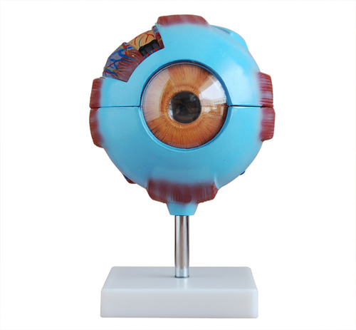
Tell us about your requirement

Price:
Quantity
Select Unit
- 50
- 100
- 200
- 250
- 500
- 1000+
Additional detail
Mobile number
Email
Name
Comapny Name
Phone Number
Email Id
City / State
Confirm Your Requirement
Verification Code
Did not receive yet?
Resend OTP

You’re Done!
We have received your requirements and will reply shortly with the best price.
Products You May Like
More Products in Anatomical Models Category
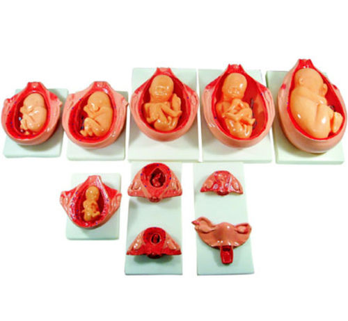
ConXport The Development Process for Fetus
Feature : Detachable, Lifelike Detailing, Stagewise Representation
Type : Fetal Development Model
Equipment Materials : Durable PVC Plastic
Dimension (L*W*H) : Varies; typically 39 x 27 x 15 cm
Core Components : Fetal Development Stages, Base Stand
Weight : Approx. 34 kg
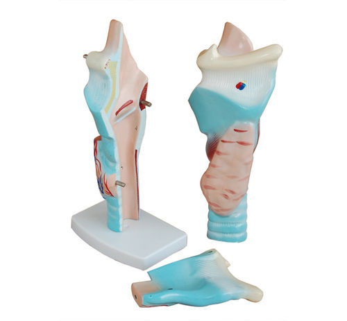
ConXport Magnified Human Larynx Model
Feature : Detailed, magnified structure for clarity
Type : Anatomical Model
Equipment Materials : Highquality PVC plastic
Dimension (L*W*H) : Approx. 25 x 15 x 13 cm
Core Components : Larynx model, removable base
Weight : Approx. 0.8 kg
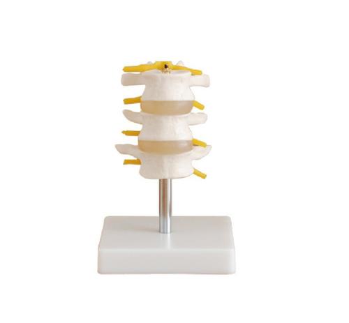
ConXport Lumbar Set (3 pcs)
Feature : Lightweight, portable, nonslip surface, washable cover
Type : Lumbar Support Blocks
Equipment Materials : Highdensity Polyurethane Foam
Dimension (L*W*H) : Standard sizes (set contains 3 different sizes)
Core Components : Three Lumbar Support Blocks
Weight : Each piece approx. 250g
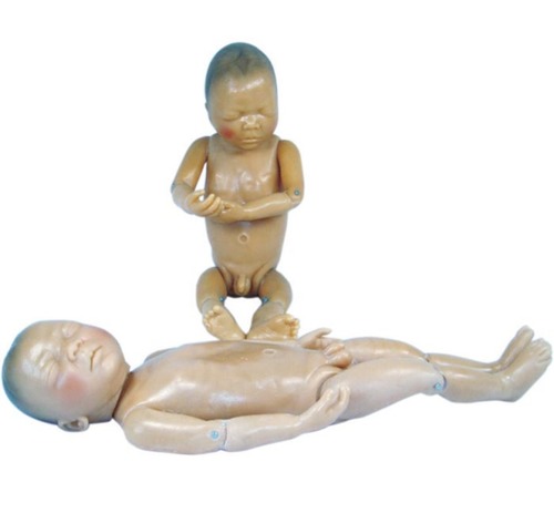
ConXport Newborn Model
Feature : Realistic Anatomical Details, Flexible Joints
Type : Physical Newborn Model
Equipment Materials : PVC (Polyvinyl Chloride)
Dimension (L*W*H) : Approx. 50 cm (Length)
Core Components : Full Body Newborn Manikin
Weight : Approx. 1.3 kg
X
×
Add a mobile number to receive call from "CONTEMPORARY EXPORT INDUSTRY"
×
Enter OTP
×
Share additional details for a quick response
×
Thank You!
Thank You for your valuable time. We have received your details and will get back to you shortly.
|
CONTEMPORARY EXPORT INDUSTRY
GST : 06AJMPT4011D1ZG
GST : 06AJMPT4011D1ZG
- B No. 4353, Main Road, Near Geeta Gopal Temple,Ambala Cantt - 133001, Haryana, India
- Phone :08045815886
- Mr Akhil Trehan (Proprietor)
- Mobile :08045815886
- Send Inquiry
Our Products
- Histopathology Products
- Projector
- Microtomes
- Pharmacy Equipment
- Covid-19 Products
- Ambulance And Recovery Equipment
- Anesthesia Equipment
- Autopsy Equipment
- Infant Care Equipment
- Blood Bank Equipments
- Diagnostic Products
- Hospital Furniture
- Hospital Hollowares
- Weight And Measuring Scale
- ICU Products
- Hospital Disposables
- X-Ray Products
- Medical Kits
- Hospital Rubber Products
- Hospital Safety Products
- OT Lights
- OT Tables
- Rehabilitation Equipments
- Suction Machines
- Surgical Instruments
- Centrifuge Machine
- Hospital Room Setup Package
- Autoclave
- Oxygen Setup
- Veterinary Instruments
- Hospital Garments
- Physiotherapy Product
- Aerosol Disinfector
- GEL ICE PACK FOR VACCINE
- Cold box
- Digital Audiometer Decent
- Audiometer
- BP Meter
- Microscopes And Projector
- Microscope
- Anatomical Models
- ENT And Ophthalmic Instruments
- Magnifying Glass Folding
- Analytical Equipment
- PCR Machine
- Analysers
- Spectrophotometer
- Photo Colorimeters
- Cold Chain Equipment
- Pathology Laboratory Equipment
- Laboratory Instruments

CONTEMPORARY EXPORT INDUSTRY
All Rights Reserved.(Terms of Use)
Developed and Managed by Infocom Network Private Limited.
Developed and Managed by Infocom Network Private Limited.

